What Does Your Blast Count Say About Your Immune System?

Disclaimer: This article is for informational purposes only and is not intended to diagnose any conditions. LifeDNA does not provide diagnostic services for any conditions mentioned in this or any other article. 

What are Blast Cells?

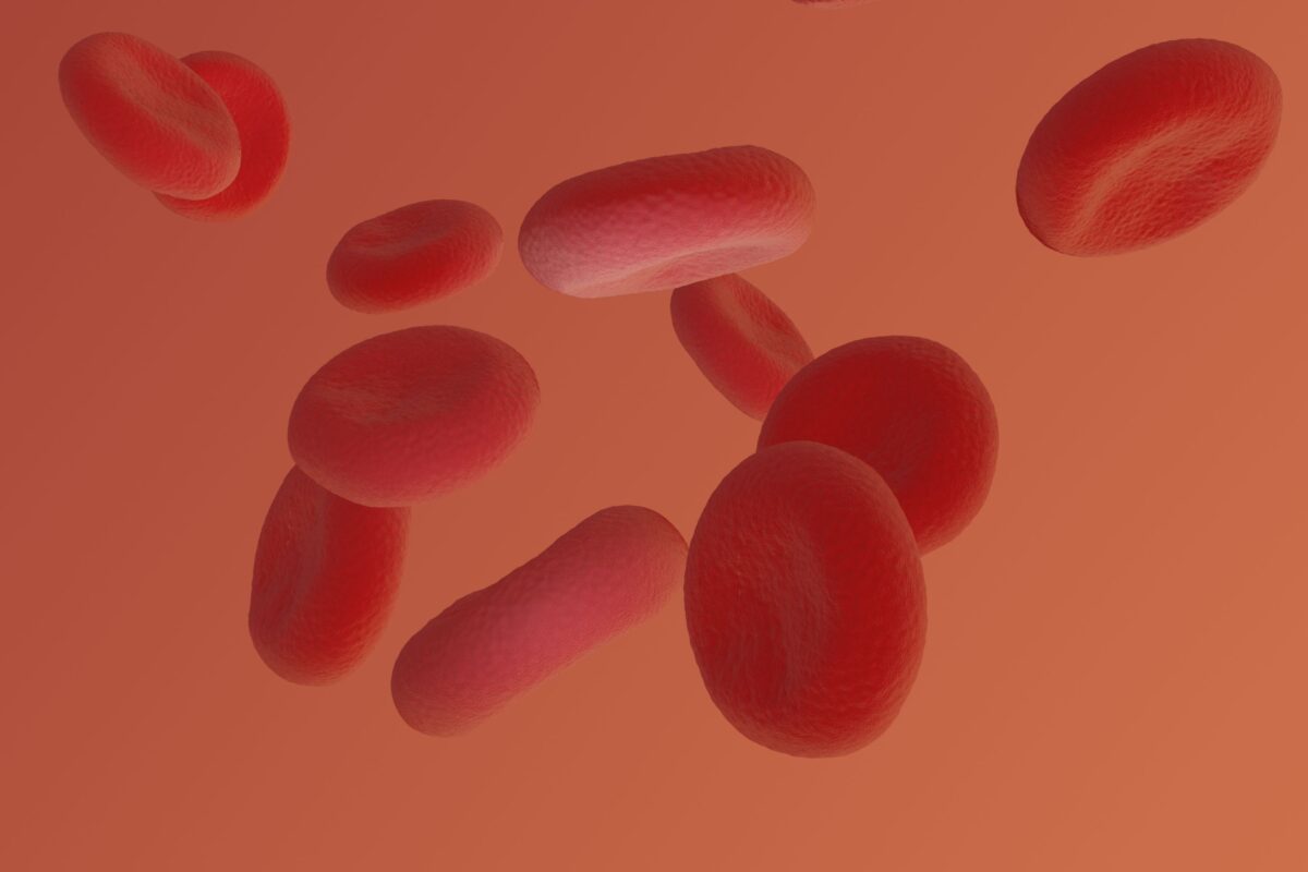
Blast cells are immature blood cells found in the bone marrow, where they develop into red blood cells, white blood cells, or platelets. Blast count refers to the number of blast cells. These immature cells play a crucial role in creating new blood cells in a process called hematopoiesis, which happens continuously throughout life. Normally, blast cells stay in the bone marrow until they mature. However, in certain health conditions, they can appear in the bloodstream too early, which is a sign that something is wrong with how the bone marrow is working.

Blast cells usually make up less than 5% of the total bone marrow cells. If they appear in the marrow in larger amounts, or in the bloodstream , it could mean the bone marrow is producing too many immature cells or not maturing them properly. This can lead to problems such as anemia (low red blood cell count), a higher risk of infections, or issues with blood clotting. Conditions like myelodysplastic syndrome (MDS) and leukemia often cause blasts to flood into the bloodstream, where they normally shouldn’t be found.

Blast cells come from hematopoietic stem cells, which are the “parent” cells in the bone marrow. These stem cells develop into one of two types of cells: myeloid or lymphoid. 

There are two main types of blast cells based on the cell lineage they are destined to follow:

  • Myeloid Blasts: These immature cells develop into granulocytes (such as neutrophils, eosinophils, and basophils), monocytes, and other myeloid cells.
  • Lymphoid Blasts: These blasts mature into lymphocytes, a key part of the immune system that includes B cells, T cells, and natural killer cells.

When doctors find a high level of blast cells in the blood, it’s a red flag for serious conditions like acute myelogenous leukemia (AML) or MDS. The type of blast cells—whether they are myeloid or lymphoid—helps doctors diagnose the exact disorder and determine the best course of treatment.

What is a Blast Count?

A blast count refers to the number of immature blood cells, or blast cells, present in the bone marrow or bloodstream. This count is typically expressed as a percentage of the total white blood cells in the bone marrow or blood sample. In healthy individuals, blast cells usually make up less than 5% of the bone marrow cells and are rarely found in the blood.

Why do Blasts Matter?

Blast cells are essential for producing healthy blood cells, but their significance goes beyond their normal role in hematopoiesis. Blasts matter because they can indicate the presence of severe conditions, such as hematopoietic neoplasms, which are disorders that affect blood cell production in the bone marrow. These conditions can disrupt the normal development of blood cells, leading to various health problems.

For example, acute leukemia is one of the most dangerous hematopoietic neoplasms where blasts rapidly multiply and take over the bone marrow, crowding out healthy blood cells. Without prompt treatment, this can quickly become life-threatening. Other disorders, like myelodysplastic syndromes (MDS) and chronic myeloproliferative disorders, also feature elevated blast levels and can gradually impair the bone marrow’s ability to function properly.

Blasts can also circulate in the bloodstream due to other factors such as severe infections, certain medications (like granulocyte colony-stimulating factor), or bone marrow-replacing processes. While not always a sign of cancer, the presence of circulating blasts should always be investigated, as it can point to serious underlying conditions.

How Do You Measure Blast Count?

Blast count is assessed through either a blood test or a bone marrow biopsy, depending on the patient’s condition. Both methods provide insight into how well the bone marrow is functioning.

  1. Blood Test (CBC with Differential): A complete blood count (CBC) with differential can estimate blast count if blasts are present in the peripheral blood. Normally, blasts are not detectable in a healthy person’s blood. If found, even in small amounts, it may indicate a bone marrow issue. While less invasive, this test may not capture an accurate blast count if levels are low or confined to the marrow.
  2. Bone Marrow Biopsy: This is the most accurate method for measuring blast count. A small bone marrow sample, usually from the pelvic bone, is examined to determine the percentage of blast cells. A healthy bone marrow contains less than 5% blasts. A higher count or blasts in the bloodstream can indicate serious blood disorders like acute myelogenous leukemia (AML) or myelodysplastic syndromes (MDS).

Why Blast Count Matter

Blast count is a crucial diagnostic tool for identifying and monitoring blood disorders. In healthy individuals, blasts should remain in the bone marrow. If they appear in the bloodstream or exceed 5% in the marrow, it may signal disorders like AML or MDS, which can disrupt normal blood cell development and lead to symptoms such as fatigue, infections, or abnormal bleeding.

Tracking blast count helps doctors evaluate disease progression and treatment effectiveness. A rising count may indicate worsening disease, while a declining count could suggest treatment success. Monitoring these changes enables more informed treatment decisions.

Blasts are measured either as a percentage of white blood cells or by their number per liter of blood. Regular monitoring is vital, especially in conditions like MDS, which can progress into more serious diseases.

What is the Normal Blast Count?

The normal blast count in healthy individuals typically comprises less than 5% of the total cells in the bone marrow. In peripheral blood, blasts should be zero or found in very low numbers.

What Does it Mean if You Have High/Low Blast Count?

High Blast Count

An elevated blast count can signal several health issues:

  • Leukemia: High blast counts are commonly associated with leukemia, a cancer that impacts blood and bone marrow. The specific type of leukemia, such as acute myeloid leukemia (AML) or acute lymphoblastic leukemia (ALL), can often be identified based on the characteristics of the blast cells.
  • Bone Marrow Disorders: Conditions like myelodysplastic syndromes (MDS) can lead to increased blast counts as the marrow struggles to produce mature blood cells.
  • Other Malignancies: Certain cancers can cause secondary increases in blast counts due to their effects on the bone marrow.

To diagnose acute leukemia, criteria include having 20% or more blasts in the peripheral blood or bone marrow, or the presence of specific leukemia gene mutations.

Types of Leukemia and Their Characteristics

  • Acute Promyelocytic Leukemia (APL): Recognized for its association with disseminated intravascular coagulation (DIC) and its unique treatment with all-trans retinoic acid (ATRA). Blasts in APL are large, have abundant cytoplasm, and display distinctive bilobed nuclei.
  • Acute Monocytic Leukemia: Characterized by leukocytosis and monocytosis, with variable blast counts. Diagnosis requires 20% blasts or promonocytes in the blood or marrow.
  • Lymphoblastic Leukemia: Lymphoblasts are small to medium-sized with scant cytoplasm and immature nuclei. Distinguishing lymphoblasts from lymphocytes can be challenging, often requiring flow cytometry.

High blast counts can indicate serious conditions, and monitoring these levels is essential for effective diagnosis and treatment planning.

Low Blast Count

A low or undetectable blast count in the peripheral blood or bone marrow generally indicates a healthy state. However, very low counts may suggest that the bone marrow is under severe stress or not producing enough blood cells.

In the context of leukemia, the presence of blasts in the blood is a crucial indicator. If more than 20% of cells in the blood are blasts, it likely points to leukemia. However, a lower percentage may occur if cancerous cells are trapped in the bone marrow, making them undetectable in blood tests.

Patients with leukemia may present with extremely high white blood cell counts, sometimes reaching between 100,000 to 400,000 per microliter of blood. Conversely, some may have low counts if immature cells are retained in the marrow.

A decreasing number of blasts typically indicates a positive response to treatment, while a rising count can signal a potential relapse.

What Indicates Remission?

Remission can vary based on individual circumstances. Two common categories include complete remission and complete remission with incomplete hematologic recovery. A patient may be considered in complete remission if they:

  • No longer require regular transfusions
  • Have a hemoglobin count that, while lower than normal, is above 7
  • Show no blasts in the blood
  • Maintain a platelet count over 100,000 (but below the normal range of 150,000)
  • Have a neutrophil count exceeding 1,000

Monitoring these parameters is essential for determining remission status and guiding ongoing treatment.

The Role of Blast Count in MDS and AML: Insights from Genetic Factors

Blast count is a critical factor in the classification and treatment of myelodysplastic syndromes (MDS) and acute myeloid leukemia (AML). Recent studies have revealed the intricate relationship between blast percentages and genetic mutations, highlighting how these elements together impact prognosis and treatment strategies.

In a 2023 Study, researchers established a clear relationship between blast count and overall survival. Higher blast percentages generally correlated with poorer outcomes. However, the presence of certain genetic mutations, such as those affecting genes TP53 or FLT3 (a gene that produces a protein that helps form and grow new blood cells), could offer better prognostic information even in patients with elevated blast counts. This finding suggests that while blast count is essential, incorporating genetic profiling enhances the understanding of patient prognosis.

Another recent study focused on the interactions between blast count and specific mutations in MDS. For instance, patients with lower blast counts who also have the SF3B1 mutation demonstrated significantly better survival rates compared to those with higher blasts. This highlights the importance of genetic factors—such as the presence of SF3B1 mutations—in influencing outcomes, thereby suggesting that assessments should include both blast percentage and genetic mutation status for a more accurate prognosis.

Clearly, the relationship between blast count and genetic factors is important for managing MDS and AML. While blast percentage is a key part of classification, it’s evident that including genetic information—like mutations in genes TP53,  FLT3-ITD and SF3B1—can greatly improve prognosis and treatment plans. 

Summary

  • Blast cells are immature blood cells in the bone marrow that develop into red and white blood cells or platelets.
  • Blast cellsplay a vital role in continuous blood cell production through a process called hematopoiesis.
  • Normally, blast cells stay in the bone marrow until they mature and make up less than 5% of total cells there.
  • If blast cells appear in the bloodstream this indicates potential issues with bone marrow function.
  • Increased blast cells are associated with  health problems like anemia, infections, and bleeding disorders.
  • There are two main types of blast cells: myeloid blasts and lymphoid blasts.
  • Myeloid blasts develop into various white myeloid blood cells, while lymphoid blasts mature into lymphocytes..
  • A blast count measures the number of immature cells in the blood or bone marrow, expressed as a percentage.
  • A normal blast count is less than 5% in the bone marrow and ideally zero in the blood.
  • High blast counts often signal serious conditions like leukemia or myelodysplastic syndromes (MDS).
  • Tracking blast count changes helps assess disease progression and treatment effectiveness.
  • An elevated blast count, particularly over 20%, typically indicates leukemia.
  • A low or absent blast count usually suggests healthy bone marrow, but very low counts may indicate systemic stress or inadequate blood cell production.
  • Remission is assessed by the absence of blasts in the blood and stable blood cell counts.
  • Genetic factors play a significant role in how blast counts affect prognosis and treatment strategies.
  • Recent studies indicate that certain genetic mutations can influence survival rates in patients with MDS and acute myeloid leukemia (AML).
  • Tailored treatment approaches are necessary as responses to therapies can differ between older and younger patients.
  • Understanding both blast counts and genetic information is crucial for effective management of blood disorders.
  • Proper monitoring can enhance patient outcomes and inform treatment decisions.
  • Recent advancements in genetic testing may allow clinicians to predict patient outcomes more accurately, making personalized therapies important in treating blood cancers like MDS and AML. 
  • Integrating genetic profiling with blast count analysis helps refine prognosis, ensuring more targeted and effective treatments that improve long-term survival and disease management.

References:

  1. https://www.verywellhealth.com/overview-of-blast-cells-4114662
  2. https://www.corpath.net/blasts
  3. https://www.biron.com/en/glossary/blast-ratio-blast/
  4. https://www.healthline.com/health/leukemia/leukemia-white-blood-cell-count-range#outlook
  5. https://www.nature.com/articles/s41375-023-01855-7
  6. https://www.ncbi.nlm.nih.gov/pmc/articles/PMC5486407/ 

Can Genes Influence Your Metamyelocyte Count

Disclaimer: This article is for informational purposes only and is not intended to diagnose any conditions. LifeDNA does not provide diagnostic services for any conditions mentioned in this or any other article.

Have you ever received a blood test result that included a term you didn’t recognize, like “metamyelocyte count”? While it may sound intimidating, understanding this may provide valuable insights into your overall health. Recognizing its significance may help you gain insight into your body’s response to stress and disease, which may potentially lead to more informed decisions about your health care.

What is Metamyelocyte Count?

Metamyelocytes are immature neutrophils in the early stages of white blood cell development, which play an important role in your immune system by helping your body fight infections. In a healthy individual, metamyelocytes are typically confined to the bone marrow where blood cells are produced and do not appear in the bloodstream. However, under certain conditions—such as infection, inflammation, or stress—the body may release immature white blood cells, including metamyelocytes, into the bloodstream.

The metamyelocyte count refers to the number of these immature cells found in a blood sample. In a routine blood test, metamyelocytes are not usually present in the blood. If they are detected, it might suggest that the bone marrow is producing white blood cells rapidly in response to an issue, such as an infection or another underlying condition. Doctors use the metamyelocyte count as part of a broader blood test to help diagnose and monitor various health conditions.

How Do You Measure Metamyelocyte Count?


The metamyelocyte count is measured as part of a routine blood test called a complete blood count (CBC) with a differential (including a breakdown of different white blood cell types). This test provides doctors with a detailed overview of your blood’s composition, particularly focusing on the various types of cells present, including those involved in your immune response.


Here’s what you can expect if your doctor orders this test:


  • Getting a Blood Sample: A small amount of blood is drawn, usually from a vein in your arm. This process is quick and typically done during a visit to your doctor or at a lab.
  • Analyzing the Blood: Once your blood sample reaches the lab, it iss analyzed to determine the types and quantities of cells present. The lab specifically looks for any immature white blood cells, such as metamyelocytes, which are not normally found in circulating blood.
  • Understanding the Results: Normally, metamyelocytes are only found in the bone marrow, not in the bloodstream. If they show up in your blood, it could be a sign that your body is responding to a particular condition, such as an infection, inflammation, or another type of stress.
  • What the Results Mean for You: If your metamyelocyte count is higher than normal, your doctor might look into other tests or ask about symptoms you’re experiencing. This helps them figure out if there’s an underlying condition that needs attention.

What is the Normal Metamyelocyte Count?

While genetic factors play a role in the normal proportions of various blood cells, the specific genetic variants  influencing metamyelocyte counts are not well-defined. However,  the normal levels of metamyelocytes may vary. While their presence in the bloodstream is generally abnormal, their presence in the bone marrow is part of the normal blood cell maturation. Understanding this helps in assessing whether their presence indicates a health issue.

Presence in Blood

The normal metamyelocyte count is zero when measured in the bloodstream. If metamyelocytes are detected in the bloodstream, it is generally considered abnormal and may indicate that the body is responding to an underlying condition. The presence of metamyelocytes in the blood may need further examination to determine the cause and assess the need for treatment.

In Bone Marrow

In the bone marrow, metamyelocytes are a normal part of the development process for white blood cells. While there isn’t a specific “normal” count for metamyelocytes in the bone marrow due to variations in individual health and age, their presence is expected as they mature into more specialized white blood cells. The bone marrow typically contains a range of immature white blood cells, including metamyelocytes, which are part of the normal hematopoiesis (blood cell formation) process.

What Causes High or Low Metamyelocyte Count?

High Metamyelocyte Count

A high metamyelocyte count means that there are more of these immature white blood cells in your bloodstream than usual. This is not typical, as metamyelocytes should normally only be found in the bone marrow. Elevated levels can suggest several potential issues:

  • Infection: The body may release immature white blood cells into the bloodstream while fighting an infection.
  • Inflammation: Chronic inflammation or conditions causing ongoing stress to the body may lead to increased metamyelocyte production.
  • Bone Marrow Disorders: Conditions like chronic myelogenous leukemia (CML), myelodysplastic syndromes (MDS), or other myeloproliferative disorders may lead to an increased release of immature cells, including metamyelocytes, into the bloodstream.
  • Severe Physical Stress: Physical stress, severe illness, or trauma can also cause a temporary increase in metamyelocyte count as the body mobilizes its immune defenses.
  • Toxic Exposure: Exposure to certain toxins, chemotherapy, or radiation may damage the bone marrow, leading to an increased release of immature cells.

Low Metamyelocyte Count

A low metamyelocyte count is rarely discussed because metamyelocytes are typically not present in the bloodstream under normal conditions. Their presence is expected primarily in the bone marrow, where they develop as part of the neutrophil maturation process. However, if their count is unusually low in the bone marrow, it may signal an underlying issue affecting bone marrow function.

For example, aplasia or dysfunction of the bone marrow can impair the production of metamyelocytes and other essential blood cells.

  • Aplastic Anemia: Aplastic anemia is a serious condition in which the bone marrow fails to produce enough blood cells, including white blood cells (WBCs) and their precursors, such as metamyelocytes. In this scenario, a low metamyelocyte count reflects the marrow’s inability to generate and release sufficient immune cells. This deficit weakens the body’s immune response, leaving it vulnerable to infections and other complications.
  • Certain Types of Leukemia: In some forms of leukemia, particularly those where the bone marrow becomes crowded with abnormal, cancerous cells, the production of normal blood cells is significantly hindered. As a result, the number of healthy precursor cells, including metamyelocytes, may be lower than expected. A bone marrow biopsy in these cases often reveals a diminished count of metamyelocytes, indicating that the marrow’s capacity to produce normal blood cells is compromised.
  • Chronic Diseases: Long-standing chronic illnesses, such as autoimmune diseases, chronic infections, or severe inflammatory disorders, can lead to bone marrow suppression. This results in a reduced production of all blood cell types, including neutrophil precursors like metamyelocytes.
  • Malnutrition: Nutritional deficiencies, particularly those affecting vitamins and minerals essential for blood cell production (e.g., vitamin B12, folate, or iron), can impair the bone marrow’s ability to produce white blood cells. In cases of severe malnutrition, a bone marrow examination may reveal fewer immature cells, including metamyelocytes.

When Should Metamyelocytes Be Detected in Blood?

Metamyelocytes should not typically be present in peripheral blood. However, they may appear in the bloodstream under certain conditions, such as:

  • Severe Infections: When the body requires an urgent response to infection, the bone marrow may release immature cells like metamyelocytes into the blood.
  • Hematologic Disorders: Conditions affecting the bone marrow, such as leukemia or myeloproliferative disorders, may result in the release of metamyelocytes into circulation.

Immature Neutrophils as Indicators of Sepsis 

Myelocytes and metamyelocytes, as well as other immature neutrophils are emerging as important biomarkers for assessing infection severity and predicting outcomes. Band cells, another type of immature neutrophil, signal severe infections like sepsis when present in high numbers—a condition known as a “shift to the left.” Elevated band cell levels are linked to poorer sepsis outcomes. 

Research has demonstrated that high percentages of band cells are associated with poorer outcomes in sepsis. Specifically, a groundbreaking study found that band cells had a sensitivity of 84% and a specificity of 71% for detecting definite sepsis at an 8.5% cutoff point. This makes band cells a valuable tool for differentiating between sepsis and non-infectious systemic inflammatory response syndrome (N-I SIRS).

Myelocytes and metamyelocytes, though less commonly monitored, also provide critical prognostic information. The same study discovered that patients with up to 40% of these cells in their blood had significantly higher mortality rates. Specifically, patients who died within the first week of ICU stay had a median of 9% myelocytes and metamyelocytes in their blood, compared to much lower levels in those who survived longer.

The presence of immature neutrophils in the blood, particularly in ICU settings, is associated with higher mortality risk, highlighting their potential as valuable markers in managing severe infections. Monitoring these cells aids in distinguishing between systemic inflammatory response syndrome (SIRS) and sepsis, thus guiding treatment and improving patient management.

Non-Genetic Factors Influencing Metamyelocyte Count

Several non-genetic factors can influence the metamyelocyte count in your blood. These factors often relate to the body’s response to various conditions or stressors. Here’s how they might affect your metamyelocyte levels:

  • Infections: During infections, especially the severe ones with bacterial origin like sepsis, the body releases immature white blood cells, such as metamyelocytes, into the bloodstream. This response is part of the immune system’s effort to rapidly combat the invading pathogens.
  • Inflammation: Chronic inflammation or inflammatory diseases such as rheumatoid arthritis or inflammatory bowel disease, can stimulate the bone marrow to produce more white blood cells, including metamyelocytes, as the body attempts to manage and repair affected tissues.
  • Stress: Physical or emotional stress can lead to an increase in the production of white blood cells, including metamyelocytes, as part of the body’s heightened immune response. For example, severe trauma or surgery can temporarily elevate metamyelocyte counts.
  • Bone Marrow Disorders: Disorders affecting the bone marrow, such as leukemia or myelodysplastic syndromes, can result in the abnormal production and release of immature cells like metamyelocytes into the bloodstream. In acute myeloid leukemia (AML), this process is particularly pronounced, leading to elevated metamyelocyte levels.
  • Medications: Certain medications, such as granulocyte colony-stimulating factor (G-CSF) used during chemotherapy, can stimulate bone marrow activity and increase metamyelocyte counts as they enhance white blood cell production.
  • Nutritional Deficiencies: While nutritional deficiencies are less directly linked to metamyelocyte counts, they can impact overall blood cell health. Deficiencies in essential nutrients like vitamin B12 or folate may indirectly affect white blood cell production by impairing bone marrow function.
  • Systemic Diseases: Systemic diseases, such as chronic kidney disease or liver disease, can alter bone marrow function and lead to changes in blood cell counts, including metamyelocytes. Chronic kidney disease, for instance, may influence the production of immature cells in the blood.


Summary


  • Metamyelocytes are immature white blood cells in early stages of development, maturing into neutrophils which play a crucial role in the immune system by fighting infections.
  • These immature cells are normally found only in the bone marrow.
  • The metamyelocyte count is assessed via a complete blood count (CBC) with a differential, which provides a detailed breakdown of different white blood cell types. Typically, metamyelocytes are absent from the bloodstream.
  • In a healthy individual, the normal metamyelocyte count in the blood is zero. Their presence in the bloodstream suggests a potential health issue and may necessitate further investigation.
  • In the bone marrow, metamyelocytes are a normal part of the blood cell maturation process, with no specific “normal” count due to variability among individuals.
  • A high metamyelocyte count can indicate various issues, including infections, chronic inflammation, bone marrow disorders, severe physical stress, or toxic exposure. Elevated levels, especially in blood,  are important for diagnosing and monitoring health conditions.
  • A low metamyelocyte count is less frequently discussed but may suggest bone marrow problems or chronic diseases affecting white blood cell production.
  • Metamyelocytes, along with other immature neutrophils like band cells, are used to assess the severity of infections and patient prognosis. High levels of band cells often signal severe infections such as sepsis and can differentiate between sepsis and non-infectious systemic inflammatory response syndrome (N-I SIRS).
  • Elevated myelocyte and metamyelocyte counts are associated with poorer outcomes in ICU settings and can help predict patient prognosis. High levels correlate with a higher risk of mortality.

References

  1. https://imagebank.hematology.org/image/60398/metamyelocyte 
  2. https://www.sciencedirect.com/topics/immunology-and-microbiology/metamyelocyte 
  3. https://www.sciencedirect.com/topics/agricultural-and-biological-sciences/metamyelocyte 
  4. https://www.biron.com/en/glossary/metamyelocyte/ 
  5. https://www.ncbi.nlm.nih.gov/pmc/articles/PMC4355545/
  6. https://www.ncbi.nlm.nih.gov/pmc/articles/PMC8081893/#B79
  7. https://karger.com/mpp/article-abstract/16/5/344/203779/The-Diagnostic-Value-of-Absolute-Neutrophil-Count 


The Role of Bilirubin in Your Health

Disclaimer: This article is for informational purposes only and is not intended to diagnose any conditions. LifeDNA does not provide diagnostic services for any conditions mentioned in this or any other article.

Bilirubin is a yellow substance produced when your body breaks down old red blood cells. This is a normal process that helps recycle iron and keep your cells healthy. Initially, bilirubin is in an unconjugated form, which means it’s not ready to be removed from the body. It then travels to the liver, where bilirubin is transformed into a form that’s easier for the body to get rid of. This process makes bilirubin conjugated and water-soluble, so it can be expelled through bile and eventually leave the body in stool. A healthy liver handles most of the bilirubin by releasing it into the bloodstream, where it’s carried to the intestines to be excreted.

Understanding bilirubin and regularly monitoring its levels is essential for evaluating liver function and overall health. By keeping track of bilirubin levels, healthcare providers may detect potential issues early on, making sure to take effective treatment. 

You may also like: Does Genetics Influence Liver Enzyme Levels?

What Does Bilirubin Level Indicate?

Bilirubin levels in the body provide important insights into overall health and may indicate various underlying health issues. Abnormal bilirubin levels may suggest different health problems, which can be grouped into causes and consequences:

Causes of High Bilirubin Levels:

  • Liver Function: Bilirubin is processed in the liver, where it becomes water-soluble and ready for excretion. If the liver is damaged or not functioning properly, bilirubin can accumulate in the blood, leading to elevated levels.
  • Hemolytic Anemia: Conditions like hemolytic anemia cause red blood cells to break down faster than normal, resulting in higher levels of unconjugated bilirubin in the blood.
  • Bile Duct Obstruction: Blockages in the bile ducts, such as gallstones or tumors, can prevent bilirubin from being excreted, leading to a buildup in the blood.
  • Hepatitis: Hepatitis, an inflammation of the liver often caused by a viral infection, hinders the liver’s ability to process bilirubin, resulting in higher blood levels.
  • Intrahepatic Cholestasis of Pregnancy: This condition, occurring during the last trimester of pregnancy, slows or stops bile drainage from the liver, impairing bilirubin processing and leading to increased bilirubin levels.

Consequences of High Bilirubin Levels:

  1. Jaundice: When bilirubin levels become too high, it can lead to jaundice, where the skin and eyes take on a yellowish hue. This condition can occur in both adults and newborns. In newborns, it may cause neonatal jaundice, which often resolves on its own but might require treatment if levels are very high.
  2. Gallstone Formation: Persistently high levels of bilirubin can contribute to the formation of gallstones, where substances like bilirubin or cholesterol harden in the gallbladder.

What is the Normal Range of Bilirubin?

In adults, typical bilirubin levels range from 0.2 to 1.2 milligrams per deciliter (mg/dL). Levels above 1.2 mg/dL are generally considered elevated and may indicate underlying health issues. For children between the ages of 15 days and 18 years, healthy bilirubin levels should be below 1.0 mg/dL.a=A nomogram can be used to assess the risk of hyperbilirubinemia (high bilirubin levels) in newborns based on their postnatal age in hours. For newborns, the interpretation of bilirubin levels depends on specific conditions and the rate of increase. For example, a bilirubin level of 8 mg/dL on the second day of life might not be alarming on its own. However, if the level was 3 mg/dL on the first day, a rapid increase to 9 mg/dL could signal the need for closer monitoring and possible intervention. 

Genetic Factors Influencing Bilirubin Levels

Bilirubin levels in the blood can significantly vary due to various factors, including genetics. Multiple studies (Telega, 2018; Grossman, 2011; Chowdhury, 2012) have identified a gene linked to Gilbert’s Syndrome (GS), which is inherited within families, and causes high blood bilirubin levels. Gilbert’s Syndrome is a common and harmless genetic condition that affects up to 8% of the population. It is inherited in an autosomal dominant manner, meaning that if one parent possesses the gene mutation, their children have a possibility of inheriting it as well. The condition is more frequently seen in boys than girls and typically appears in older children or teenagers.

GS is caused by a mutation in the UGT1A1 gene. This gene helps produce an enzyme needed to convert bilirubin, a substance made by the liver, into a form that can be easily removed from the body. In people with GS, this enzyme doesn’t work as well, leading to higher levels of bilirubin in the blood.

Despite the biochemical changes, around one-third of individuals with GS show no symptoms, and the condition is often discovered accidentally during blood tests conducted for other reasons. When symptoms do occur, they typically involve mild, persistent, or occasional yellowing of the skin and eyes (jaundice), especially during periods of stress, illness, fasting, or alcohol consumption.

Doctors diagnose GS by noting mild increases in bilirubin levels without signs of liver damage or red blood cell breakdown. Blood tests usually show normal liver function. The diagnosis can be confirmed if bilirubin levels increase significantly after a 24-hour fast. Genetic testing can also identify the specific UGT1A1 gene mutation responsible for the condition.

Interestingly, GS might have some health benefits. Research by Kundur et al. (2015) has shown that people with GS have higher levels of antioxidants in their blood, including bilirubin and glutathione. These antioxidants defend the body against damage caused by harmful molecules related to  oxidative stress.

The study suggests that individuals with GS have a decreased risk of heart disease and associated deaths. This protection might be due to elevated bilirubin reducing the activity of platelets, the small blood cells that form clots. Oxidative stress can enhance platelet activity, which raises the risk of blood clots and heart attacks. By acting as a natural anti-clotting agent, bilirubin may lower the risk of heart attacks and explain why people with GS have lower heart disease mortality.

Some FAQs on Bilirubin

What is a Bilirubin test?

A bilirubin test checks the amount of bilirubin in your blood to help doctors evaluate your liver performance. It is commonly included in a group of tests that assess liver health. Normally, a healthy liver processes and removes bilirubin efficiently. However, if you have liver problems, bilirubin can accumulate to unhealthy levels in your body, indicating potential liver disease.

Why do I need a bilirubin blood test?

Doctors frequently request a bilirubin blood test to diagnose and monitor different health conditions.

You might require this test if you have symptoms like yellowing of the skin or eyes, dark-colored urine, or stomach pain, as these could be signs of liver issues such as hepatitis, cirrhosis, or gallbladder problems.

The test can also help determine if there’s a blockage in the bile ducts. For individuals with existing liver diseases or disorders, a bilirubin test is essential for monitoring the condition and assessing liver function. In some cases, concerns related to the breakdown of red blood cells, such as in cases of hemolytic anemia, occur. 

Why do many newborns develop jaundice, and how is it treated?

Many newborns develop jaundice a few days after birth due to high bilirubin levels. This happens because newborns have more red blood cells that break down quickly, but their livers are not yet fully developed to process bilirubin efficiently. Before birth, the pregnant person’s liver helps manage bilirubin. A newborn’s liver is only about 1% as active as an adult’s liver.

In most newborns, jaundice appears within 1 to 3 days after birth, with bilirubin levels peaking around the fourth or fifth day. These levels can reach up to 18 mg/dL. Typically, the jaundice resolves within 2 weeks as the baby’s liver matures and becomes better at processing bilirubin.

To help reduce bilirubin levels, feeding the baby 8 to 12 times per day promotes regular bowel movements, which aids in removing bilirubin from the body. In most cases, this condition is temporary and clears up on its own. However, if jaundice persists or bilirubin levels become very high, medical treatments such as phototherapy, intravenous immunoglobulin, or exchange transfusion may be necessary.

What are the symptoms of high bilirubin?

The symptoms of high bilirubin can vary depending on the underlying cause. Some people with mildly elevated bilirubin levels may not experience any symptoms at all.

For those with moderately high bilirubin, the primary symptom is often jaundice, which is characterized by a yellowish color in the eyes and skin. Jaundice is the most common and noticeable sign of elevated bilirubin levels.

Other general symptoms that might accompany conditions causing high bilirubin include:

  • Abdominal pain or swelling: Liver or gallbladder disorders that increase bilirubin levels can cause abdominal pain or swelling due to inflammation, obstruction, or damage to these organs.
  • Chills: Chills can occur with infections or inflammation of the liver (like hepatitis), which can also raise bilirubin levels.
  • Fever: Fever may indicate an underlying infection or inflammatory condition affecting the liver, such as hepatitis or a bile duct infection, which can also lead to elevated bilirubin.
  • Chest pain: Though less directly related, severe liver disease or complications can cause chest pain, possibly due to referred pain or related cardiovascular stress.
  • Fatigue: High bilirubin can be a sign of liver dysfunction, leading to fatigue and weakness due to the liver’s role in energy metabolism.
  • Lightheadedness: Severe liver diseases causing high bilirubin can disrupt normal metabolic processes, leading to symptoms like lightheadedness due to reduced blood flow or oxygen levels.
  • Nausea: High bilirubin can result from liver or gallbladder disease, which can also cause digestive disturbances like nausea.
  • Unusually dark urine: High bilirubin levels in the blood can lead to its excretion in the urine, causing a darker color. This is particularly associated with direct (conjugated) bilirubin which is water-soluble.

Tips to Naturally Lower Bilirubin Levels

To naturally lower your bilirubin levels and support overall liver health, consider implementing the following lifestyle and dietary changes:

Eat a Balanced Diet: A diet rich in fruits, vegetables, and whole grains provides essential nutrients that support liver health. Vitamins, minerals, and antioxidants support liver function, which helps in a more efficient processing and elimination of bilirubin.

Stay Hydrated: Drink lots of water to maintain general liver health. It helps in the elimination of toxins from the body, including excess bilirubin. 

Fiber-Rich Food: Food items with high fiber like oats, beans, and whole grains may help with digestion. Improved digestion can help the body remove excess bilirubin through regular bowel movements.

Control Alcohol Consumption: Drinking too much alcohol may harm the liver’s ability to process and eliminate bilirubin. 

Refrain from Consuming Processed and High-Fat Foods: Consuming certain food itemsfood like these can increase the risk of liver inflammation and fatty liver disease. Avoiding fried foods, junk food, and processed snacks can help prevent liver damage and support healthy bilirubin levels.

Regular Exercise: Exercise regularly because it enhances blood circulation, helps to lose weight, and improves overall liver function. Physical activity reduces fat buildup in the liver and promotes better metabolic health, which can assist in lowering bilirubin levels.

Maintain a Healthy Weight: Maintain a healthy body weight to prevent liver problems. A healthy liver is more efficient at processing and eliminating bilirubin, which helps keep its levels in the normal range.

Avoid Excessive Use of Over-the-Counter Medications: Over-the-counter pain relievers, especially those containing acetaminophen, can strain the liver when used excessively. Managing medication use and consulting with a healthcare professional can help avoid liver complications.

Manage Stress: Chronic stress can impact liver function and overall health. Incorporate stress management techniques such as meditation, deep breathing exercises, and relaxation techniques to support liver health.

Limit Exposure to Toxins: Minimize exposure to environmental toxins and chemicals that can stress the liver. This includes avoiding cigarette smoke, limiting exposure to industrial chemicals, and using natural cleaning products.

References

  1. https://medlineplus.gov/lab-tests/bilirubin-blood-test/ 
  2. https://www.urmc.rochester.edu/encyclopedia/content.aspx?contenttypeid=167&contentid=total_bilirubin_blood 
  3. https://www.medicalnewstoday.com/articles/315086#in-newborns 
  4. https://www.bioma.me/bioma-health/biomarkers/the-role-of-bilirubin-in-the-body 
  5. https://www.healthline.com/health/high-bilirubin#bilirubin-levels 
  6. https://www.sciencedirect.com/topics/nursing-and-health-professions/gilbert-disease#:~:text=A%20presumptive%20diagnosis%20of%20Gilbert,postprandial%20bile%20acids%20are%20normal
  7. https://gracelaboratory.com/how-to-reduce-direct-bilirubin/
  8. https://www.sciencedirect.com/science/article/abs/pii/S0021915014016645

Why Do We Have Fear of Heights?

Disclaimer: This article is for informational purposes only and is not intended to diagnose any conditions. LifeDNA does not provide diagnostic services for any conditions mentioned in this or any other article.

Overview

Is the fear of heights something we’re born with or something we learn? This question has intrigued researchers for years. The fear of heights, also known as acrophobia, is a common phobia that affects 1 in 20 people. It may range from mild anxiety when looking down from a high place to intense panic and avoidance of any situation involving heights. 

For those affected, acrophobia may significantly impact daily life, limiting activities and opportunities. Therefore, understanding the reasons behind this fear is important for developing better strategies to manage and overcome it. By gaining insight into the genetic and non-genetic factors contributing to acrophobia, individuals and healthcare professionals can develop effective treatments improving the quality of life for those who suffer from this condition.

What is Acrophobia?

Acrophobia is a severe fear of heights that can provoke intense anxiety and panic attacks. It can develop at any stage of life but often begins in childhood or adolescence and affects both genders, though studies indicate it might be more common in women. This specific phobia involves an extreme and irrational fear of heights, separate from a general fear of falling. 

People with acrophobia may avoid activities like climbing ladders, standing on balconies, or driving over bridges, which can greatly affect their daily lives. When faced with heights, they might experience various symptoms such as dizziness, trembling, rapid heartbeat, shortness of breath, and nausea. In severe cases, even thinking about heights can cause these reactions, showing how deeply acrophobia can impact a person’s life.

What Are the Symptoms of Acrophobia?

Acrophobia is characterized by both psychological and physical symptoms, each contributing to the intense fear of heights.

Psychologically, acrophobia involves intense fear and anxiety when thinking about, looking at, or being in high places. Individuals with this phobia often worry excessively about negative outcomes in such situations, such as falling or being trapped. This fear can be so overwhelming that it leads to a strong desire to escape if the affected persons find themselves in a high place, severely impacting their ability to function normally in everyday activities involving heights.

Physically, acrophobia can trigger a range of symptoms when exposed to heights. Common physical reactions include sweating, rapid heartbeat, dizziness, and lightheadedness. Those affected may also feel queasy, tremble, and experience shortness of breath. These physical symptoms not only heighten the overall sense of fear and panic but also make it challenging to manage the condition, as the body’s responses further reinforce the psychological distress.

Non Genetic Factors Influencing Fear of Heights

Traumatic Experiences: Experiences such as falling from a significant height or witnessing an accident that involves heights may lead to the development of acrophobia. This may create a lasting association between heights and fear, triggering anxiety and panic in similar situations in the future.

Cognitive Factors: Cognitive distortions and negative thinking patterns may also contribute to the development and maintenance of acrophobia. Individuals with acrophobia may overestimate the danger of heights and underestimate their ability to cope with the situation. These distorted thoughts may intensify fear and anxiety, making it difficult to confront and overcome the phobia. Cognitive-behavioral interventions may help address these negative thought patterns.

Learning and Conditioning: Classical and operant conditioning may play a role in the development of acrophobia. Classical conditioning involves the association of heights with a negative or traumatic experience, leading to the development of fear. Operant conditioning, on the other hand, involves the reinforcement of avoidance behaviors, which may perpetuate the phobia. Understanding these learning mechanisms may inform effective therapeutic approaches.

Lack of Exposure: Limited exposure to heights and a lack of positive experiences in high places can reinforce fear. When individuals avoid high places, they miss opportunities to gradually desensitize themselves and learn that heights are not inherently dangerous. This avoidance may  intensify their fear.

Sensory Processing Issues: Difficulties with sensory processing, such as problems with balance or spatial orientation, can exacerbate the fear of heights. Individuals with vestibular disorders or issues with depth perception may experience discomfort or dizziness in high places, which can heighten their fear and anxiety.

Stress and Anxiety: High levels of general stress and anxiety can make individuals more sensitive to potential threats, including heights. Chronic stress can lower an individual’s threshold for fear and increase overall anxiety levels, making it more challenging to cope with situations involving heights.

Physical Health Conditions: Conditions affecting balance and spatial orientation, such as vestibular disorders, can contribute to the fear of heights. When physical health issues cause symptoms like dizziness or vertigo, individuals may develop a fear of heights as a result of the discomfort or disorientation experienced in high places.

Genetic Factors Influencing Fear of Heights

In a GWAS study involving 642 participants from a genetically isolated Finnish population, researchers aimed to identify genetic factors associated with acrophobia or the fear of heights. The study utilized 575 autosomal microsatellite markers to analyze the genetic makeup of individuals with pure acrophobia as well as those with acrophobia and schizophrenia.

The results revealed several chromosomal regions with potential links to acrophobia. The strongest evidence of linkage was found on chromosome 13q21-q22, specifically at marker D13S162, which achieved a LOD score of 2.22 in individuals with pure acrophobia. A nearby SNP rs2323266, located close to the protocadherin 20 (PCDH20) gene, was previously associated with schizophrenia, although it is not within the same linkage disequilibrium block as D13S162.

Additionally, chromosome 4q28, with marker D4S2394, showed a LOD score of 2.17, and chromosome 8q24.2-q24.3, with marker D8S373, showed a LOD score of 2.09. While these regions exhibited significant LOD scores, they did not achieve genome-wide significance, and prior associations with anxiety disorders or schizophrenia were not established for these regions.

Despite these findings, the study did not identify any specific high-risk gene variants shared among the families with acrophobia. This suggests that acrophobia may be influenced by a complex array of genetic factors rather than a few predominant risk alleles.  Some regions associated with acrophobia showed connections to schizophrenia, highlighting potential genetic overlaps between these conditions. 

Another study involving 45 participants investigated how acrophobia relates to balance and spatial perception. Researchers assessed balance and perception of heights through questionnaires and tests. This study found that fear of heights is closely linked to how individuals use their vision to maintain balance, their ability to stay balanced, their comfort in certain environments, and physical symptoms like dizziness. Interestingly, general anxiety did not appear to be a major factor in most cases of acrophobia. This study highlights that acrophobia is more about sensory processing and balance than generalized anxiety, suggesting a potential focus for developing more effective treatment strategies.

How to Diagnose Fear of Heights?

To diagnose severe fear of heights, acrophobia, a person typically needs to have experienced intense and persistent fear for at least six months. Healthcare providers use criteria from the Diagnostic and Statistical Manual of Mental Disorders (DSM-5) to confirm that the fear is excessive and disproportionate, involves significant anxiety about future encounters with heights, leads to avoidance of high places, and disrupts daily life. Health providers may need to also rule out other conditions that could be causing similar symptoms.

How to Treat Fear of Heights?

Treating fear of heights has a variety of options suited to individual needs and preferences. These approaches can help individuals manage and overcome their acrophobia effectively.

Cognitive Behavioral Therapy (CBT)

CBT effectively reduces anxiety and avoidance behaviors linked to acrophobia. This therapy involves recognizing and challenging unrealistic beliefs about heights. By using cognitive restructuring and gradual exposure, individuals learn to face their fears and develop more practical and balanced thinking.

Exposure Therapy

A specific form of CBT, exposure therapy involves gradually and systematically facing heights. This method helps individuals reduce their fear and anxiety by confronting heights in a controlled setting. Repeated exposure can help people become more comfortable with heights and gradually overcome their fear.

Medication

Sometimes, medications are prescribed to help manage symptoms of acrophobia. Antidepressants and anti-anxiety drugs, like benzodiazepines, can alleviate anxiety and enhance overall functioning. Medication is typically combined with therapy for a more thorough treatment. It’s essential that these medications are prescribed by a healthcare professional.

Virtual Reality Exposure Therapy

Virtual reality (VR) has emerged as a popular tool for treating acrophobia. VR exposure therapy allows individuals to gradually face their fear in a controlled, virtual environment. For instance, someone with acrophobia might begin by viewing photos of high places and then progress to a VR headset that simulates being at various heights. This method helps people slowly and safely become accustomed to their fear using coping techniques learned alongside with a therapist. 

Vestibular Physical Therapy

Some individuals with acrophobia may co-experience issues with their vestibular system, which is responsible for balance. Vestibular physical therapy aims to strengthen this balance system and teach strategies to improve stability. By addressing balance issues, this therapy can also help reduce the fear of falling that often accompanies acrophobia.

Relaxation Techniques

Techniques such as deep breathing, progressive muscle relaxation, and guided imagery can help manage the physiological symptoms of anxiety associated with heights. These practices help reduce overall stress levels and promote a sense of calm, making it easier to confront and manage fear.

Additional Lifestyle Coping Strategies

Besides formal therapies, there are various lifestyle approaches to manage acrophobia. Techniques such as deep breathing, yoga, and visualization can help reduce stress and promote relaxation. Engaging in regular physical activity, maintaining a healthy diet, and sharing feelings with a supportive person can also be beneficial in coping with acrophobia and its effects on daily life.

Gradual Exposure 

Gradual exposure involves slowly increasing exposure to heights in a step-by-step manner. This approach helps individuals build confidence and reduce fear over time. For example, starting with viewing images of heights, progressing to standing on a low platform, and eventually advancing to higher elevations.

Professional Guidance and Consultation

Consulting with a mental health professional specializing in anxiety disorders or phobias can provide personalized treatment plans and strategies. Professionals may offer guidance on selecting the most appropriate therapies, monitoring progress, and adjusting treatment approaches as needed.

Summary

  • Acrophobia, or fear of heights, affects about 1 in 20 people and can vary from mild anxiety to severe panic.
  • The condition often emerges during childhood or adolescence, and affects both genders.
  • Acrophobia involves an irrational fear of heights, leading to avoidance of activities like climbing stairs or driving over bridges.
  • Symptoms of acrophobia include dizziness, sweating, trembling, rapid heartbeat, shortness of breath, and nausea.
  • Traumatic experiences involving heights, such as falls, can contribute to the development of acrophobia.
  • Genetic studies have identified several chromosomal regions linked to acrophobia, though no specific high-risk gene variants were found.
  • A region in chromosome 13q21-q22, specifically at marker D13S162, showed the strongest evidence of linkage to acrophobia in a GWAS study.
  • Exposure therapy, a type of CBT, involves gradual exposure to heights to help individuals desensitize and manage their fear.
  • CBT helps individuals challenge and reframe distorted thoughts about heights, reducing anxiety and avoidance behaviors.
  • Medications, such as antidepressants and anti-anxiety drugs, can support acrophobia treatment when prescribed by healthcare providers.
  • Virtual Reality (VR) exposure therapy allows individuals to confront their fear of heights in a controlled, virtual environment.
  • Vestibular physical therapy addresses balance system issues that may contribute to acrophobia, improving stability and reducing fear of falling.
  • Additional coping strategies like yoga and regular exercise can help manage acrophobia and improve overall well-being.
  • Practices like deep breathing and progressive muscle relaxation help manage anxiety symptoms related to heights.
  • Facing heights step-by-step builds confidence and reduces fear over time.
  • Consulting with a mental health expert offers personalized treatment plans and progress monitoring.
  • Focusing on the present by practicing mindfulness may help reduce height-related stress.
  • Sharing experiences and strategies with others facing similar fears may help reduce fear of height.

References

  1. https://my.clevelandclinic.org/health/diseases/21956-acrophobia-fear-of-heights
  2. https://psycnet.apa.org/record/2014-34671-006
  3. https://pubmed.ncbi.nlm.nih.gov/20821801/
  4. https://www.forbes.com/health/mind/acrophobia/
  5. https://www.verywellhealth.com/acrophobia-fear-of-heights-5093778#:~:text=Like%20all%20anxiety%20disorders%20and,of%20genetic%20and%20environmental%20factors.

What Role Do Your Genes Play in the Formation of Bunions?

Disclaimer: This article is for informational purposes only and is not intended to diagnose any conditions. LifeDNA does not provide diagnostic services for any conditions mentioned in this or any other article.

What Are Bunions?

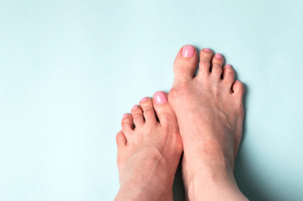
Have you ever noticed a bony bump at the base of your big toe? This common foot deformity is known as a bunion or hallux valgus. The big toe anchors your foot, connecting to the first toe bone through the metatarsophalangeal (MTP) joint, which helps distribute your weight as you walk. When this joint develops a problem, the bones can shift out of alignment, causing the big toe to lean towards the smaller toes and the joint base to protrude, often leading to red, sore skin over the area.

Genetics play a significant role in bunions, with 70% of affected individuals having family members who also experience them. While bunions may appear early in life due to genetic predispositions, they often develop during adulthood. Factors such as repetitive minor injuries from wearing high-heeled or narrow-toed shoes may contribute to their formation. Bunions are more common in adults, particularly affecting women more than men.

What Causes Bunions?

The exact reasons for bunions aren’t fully known, but experts think both inherited traits and lifestyle choices are involved. Here are the possible genetic and environmental causes for bunions:

Genetic Causes of Bunions

Inherited Factors and Foot Mechanics: Genetics may influence bunion development by shaping foot structures such as flat feet, low arches, or overly flexible joints and tendons. These inherited traits alter foot, leading to gradual misalignment of the big toe joint over time.

Congenital Deformities: Some individuals are born with foot deformities that predispose them to bunions. These congenital issues, such as abnormal bone shapes or joint structures, contribute to misalignment and bunion development from an early age.

Genetic dispositions to bunions are discussed more in detail further in this article.

Non-Genetic Causes of Bunions

Impact of Footwear: Wearing shoes that don’t fit well, particularly high heels or narrow-toed shoes, may worsen bunion risk. High heels push body weight forward, increasing pressure on the front of the foot and toes. Over time, this pressure can misalign the big toe joint and lead to bunion formation. Women are more susceptible, often due to their choice of footwear.

Arthritis and Bunion Development: Conditions like osteoarthritis and rheumatoid arthritis may contribute to bunion development by causing joint inflammation and cartilage breakdown. These factors may lead to pain, deformity, and instability in the MTP joint.

Foot Stress and Injuries: Repetitive stress or injuries to the foot, often from activities like ballet dancing, may increase the risk of bunion formation. Such injuries may destabilize the MTP joint, causing bones to shift out of place.

What Are The Symptoms of Bunions? 

The signs and symptoms of a bunion include:

Bulging Bump: A noticeable bulge at the base of the big toe is the most common symptom of a bunion. This bony bump forms as the big toe joint becomes misaligned and may vary in size and severity, depending on the extent of the misalignment.

Swelling, Redness, or Soreness: The skin over the bunion may become swollen, red, and sore due to the constant pressure from shoes. Inflammation and irritation of the surrounding soft tissues may lead to discomfort and pain, especially when walking or wearing tight footwear.

Corns or Calluses: Corns or calluses may develop on the overlapping first and second toes due to the constant rubbing and pressure. These thickened layers of skin form as a protective response to the friction and may cause additional pain and discomfort.

Ongoing Pain: The pain can range from mild to severe and may be accompanied by a burning sensation or aching in the affected joint which may worsen with prolonged standing, walking, or wearing tight shoes.

Limited Movement: The misalignment and inflammation can make it difficult to bend or straighten the toe, affecting your overall foot function which may lead to restricted movement of the big toe joint. 

Genetic Disposition to Bunions

A study involving patients and their family members with hallux valgus (HV), or bunions,  was conducted in 2007 to understand its hereditary patterns. Over a year, 1174 patients with painful bunion deformities underwent X-ray imaging and filled out detailed family history forms. The proband consisted of350 individuals (22 men, 328 women; average age 47.8 years), with a ratio of about 1 man for every 15 women. They found that in the families of 244 out of the 350 patients, three or more members had HV; in 71 families, two members were affected;and in 35 families, only the patient themselves showed symptoms. The study observed that females were more likely to have parents and relatives affected by HV. However, the severity of HV did not significantly vary based on gender or affected family members. The presence of a family history of bunions in 90% of patients and the condition appearing across multiple generations suggests that HV may be inherited dominantly but not everyone with the genetic predisposition develops symptoms.

A more recent study of 2021 involved 10 participants from three independent families, including 7 individuals affected by HV and 3 unaffected, and used whole exome sequencing, a type of DNAsequencing  currently used  to understand the genetic causes of symptoms or diseases. The DNA analysis identified significant genetic factors linked to bunions. The study identified 36 different genes potentially associated with HV, including TTN, COL6A3, and LARS, which are known for their role in influencing the development of long toes and fingers. Additionally, genes like FIG4 and CBS, which are associated with digital anomalies, were highlighted. The C7 gene which was associated with bunions is also linked to chronic arthritis, and may contribute to HV by promoting inflammation

How Are Bunions Diagnosed?

Bunions are diagnosed through a physical examination, where your doctor will look for signs such as a noticeable bump at the base of your big toe, along with swelling, redness, soreness, and limitations in toe or foot movement. X-rays are commonly used to assess the severity of the condition and the alignment of bones.

Health Impact or Complications of Bunions:

Bunions may lead to several complications if left untreated, impacting the overall health and functionality of your feet. Here are some possible complications of bunions:

Bursitis: This condition is the inflammation of the small fluid-filled pads (bursa) that cushion the bones near joints. Bunions create extra pressure and friction at the joint of the big toe. Over time, this persistent stress can irritate and inflame the bursa. The inflammation of the bursa, known as bursitis, results in pain, swelling, and restricted movement in the affected area.

Hammertoe: This occurs when the middle joint of a toe, typically the second toe, bends abnormally, often due to the pressure from a bunion altering foot mechanics. Symptoms include a visible bend, pain, and corns or calluses from friction.

Metatarsalgia: Refers to pain and inflammation in the ball of the foot, often caused by the altered weight distribution due to a bunion. This condition results in sharp or aching pain, swelling, and a sensation of walking on a pebble. 

Can You Treat Bunions? 

Treatment options vary depending on the severity of your bunion and the pain it causes. These include both non surgical and surgical methods.

Non-Surgical Options:

Nonsurgical treatments that may relieve the pain and pressure of a bunion include:

Changing shoes: Wear roomy, comfortable shoes that provide plenty of space for your toes.

Medications: Pain relief for bunions may often be managed with over-the-counter pain relievers and anti-inflammatory treatments to reduce pain and swelling. It is important to seek advice from a healthcare professional before starting any medication or treatment plan to ensure it is appropriate for your specific condition.

Shoe inserts: Padded shoe inserts may help distribute pressure evenly when you move your feet, reducing symptoms and preventing the bunion from getting worse.

Applying ice: Icing your bunion after you’ve been on your feet too long or if it becomes inflamed may help relieve soreness and swelling.

Surgical Options

If non surgical treatment doesn’t relieve your symptoms, surgery might be necessary. Surgery is not recommended for cosmetic reasons but only when a bunion causes frequent pain or interferes with daily activities. There are various surgical procedures for bunions, which can be done as single procedures or in combination. These might involve:

Osteotomy: Involves cutting and realigning the bone of the big toe. The surgeon makes small cuts in the bones to shift them into the correct position, held together with screws or pins. This procedure is often combined with other techniques to correct the toe’s alignment.

Exostectomy: Involves removing the bony bump from the toe joint without performing an alignment. While this can reduce pain and improve shoe fit, it does not address the underlying cause of the bunion and is usually combined with other procedures for long-term correction.

Arthrodesis: Involves surgical fusion of the big toe joint, creating a permanent bond between the bones. This procedure is used for severe bunions, particularly in cases where arthritis affects the toe joint. It eliminates joint movement, reduces pain and corrects deformity.

Lapidus Procedure: This procedure addresses hypermobility in the joint, by stabilizing the foot and realigning the toe. It involves fusing the first metatarsal bone to the medial cuneiform bone to correct severe bunions and prevent recurrence

Resection Arthroplasty: Involves removing the damaged portion of the toe joint to create a flexible scar tissue joint. This procedure is less common and usually reserved for older patients or those with severe arthritis and poor bone quality.

Summary

  • Bunions, or hallux valgus (HV), are bony bumps that form on the joint at the base of the big toe.
  • Both genetic factors and environmental influences contribute to the development of bunions.
  • Genetic factors are significant in the formation of bunions, with 70% of those affected having a family history of the condition.
  • Proper foot care and wearing appropriate footwear can help manage bunions and prevent them from getting worse.
  • Bunion pain can greatly affect daily activities and foot function.
  • Genes like TTN, COL6A3, LARS, FIG4, CBS, and C7 may be linked to the development of bunions.
  • Bunions are more common in adults and women, often associated with repetitive foot injuries and inflammatory conditions.
  • Symptoms of bunions include a visible bump, redness, swelling, soreness, corns, calluses, ongoing pain, and limited toe movement.
  • Inherited foot shapes, foot stress, congenital deformities, and arthritis increase the risk of developing bunions.
  • Diagnosis of bunions involves a physical examination, X-rays, and sometimes blood tests to rule out other conditions.
  • Untreated bunions can lead to complications such as bursitis, hammertoe, and metatarsalgia.
  • Surgery is considered for bunions only when they cause significant pain or interfere with daily life.
  • Preventive measures include choosing shoes with a wide toe box and adequate foot support.
  • Discovery of certain associated genes suggest that bunions may result from abnormal bone growth and chronic inflammation.
  • Understanding the genetic causes of bunions can help in early detection and targeted treatment strategies.

References

  1. https://www.lifespan.org/lifespan-living/what-are-bunions-signs-causes-treatments
  2. https://www.mayoclinic.org/diseases-conditions/bunions/symptoms-causes/syc-20354799
  3. https://www.health.harvard.edu/diseases-and-conditions/what-to-do-about-bunions
  4. https://www.mayoclinic.org/diseases-conditions/bunions/diagnosis-treatment/drc-20354805
  5. https://www.mayoclinic.org/diseases-conditions/bunions/diagnosis-treatment/drc-20354805
  6. https://orthoinfo.aaos.org/en/diseases–conditions/bunions/
  7. https://radiopaedia.org/articles/hallux-valgus
  8. https://www.mayoclinic.org/diseases-conditions/metatarsalgia/symptoms-causes/syc-20354790
  9. https://www.thefootcompany.co.uk/blog/are-bunions-genetic

What Your Finger Length Ratios Tell About Your Health and Behavior

Disclaimer: This article is for informational purposes only and is not intended to diagnose any conditions. LifeDNA does not provide diagnostic services for any conditions mentioned in this or any other article.

Overview

Have you ever wondered how something as simple as the length of your fingers could reveal insights into your health and personality? The 2D:4D ratio of finger length ratio, which compares the lengths of your index and ring fingers, is emerging as a fascinating biomarker in scientific research. It became well-known after John Manning’s research in 1998, which suggested that this ratio could show the levels of testosterone and estrogen you were exposed to in the womb.  

Studies have linked the 2D:4D ratio to various traits and behaviors, like aggression, sexual orientation, and even risks for conditions such as autism and addiction. It even helps understand links between early hormonal exposure and various outcomes, such as heart disease, mental health issues, and even personality traits. However, there’s still debate over its reliability and whether it truly reflects prenatal hormone exposure.

What Is the Finger Length Ratio?

The finger length ratio, or 2D:4D ratio, measures the length of your index finger (2D) compared to your ring finger (4D). Men usually have a lower ratio, meaning their ring fingers are longer than their index fingers. This ratio is thought to reflect the balance of testosterone and estrogen they were exposed to while in the womb. Researchers use tools like calipers and scanners to measure these ratios, exploring their links to various traits and behaviors. 

How Does the 2D:4D Ratio Relate to Health Conditions?

A recent study by researchers from Swansea University and the University of Central Lancashire explored the relationship between finger length ratios, specifically the 2D:4D ratio, and oxygen metabolism efficiency by examining 133 professional male football players. They measured the players’ finger lengths and assessed their oxygen metabolism and maximal oxygen consumption through cardiopulmonary tests on a treadmill. The findings revealed that players with a lower 2D:4D ratio, meaning longer ring fingers relative to index fingers, exhibited more efficient oxygen metabolism and higher maximal oxygen consumption. This suggests that prenatal testosterone exposure, which influences this finger ratio, may also affect mitochondrial function and energy production. 

These results align with previous research linking a lower 2D:4D ratio to better performance in distance running, reduced severity of diseases like Covid-19, and a decreased risk of heart disease. Professor John Manning emphasized the potential of the 2D:4D ratio as a marker for understanding metabolic processes related to health outcomes and called for further research to explore these associations, especially in women who were not part of the Manning study. This study adds to the growing evidence of the biological significance of finger length ratios in health and disease.

Another study titled The Ratio of Second to Fourth Digit Length (2D:4D) and Coronary Artery Disease in a Han Chinese Population explores whether the 2D:4D ratio,can serve as an indicator for susceptibility to coronary artery disease (CAD) among a Chinese population. In this study, researchers analyzed 1,764 participants, dividing them into four groups: 441 men with CAD, 441 men without CAD, 441 women with CAD, and 441 women without CAD. Finger lengths were meticulously measured using electronic calipers, and statistical methods, including receiver operating characteristic (ROC) curves, were employed to evaluate the potential diagnostic value of the 2D:4D ratio for CAD.

The results revealed that in men, the 2D:4D ratio was significantly higher among those with CAD compared to those without. Specifically, the right-hand 2D:4D ratio demonstrated a moderate diagnostic value with an area under the curve (AUC) of 0.72, suggesting that this ratio could be a useful marker for CAD risk in males. Conversely, the study did not find a significant difference in the 2D:4D ratios between women with CAD and those without, indicating that the ratio might not be a reliable indicator for CAD risk in women.

The findings align with previous research linking a higher 2D:4D ratio with increased CAD risk in men. This association is thought to be related to prenatal hormone exposure, as the 2D:4D ratio reflects hormonal influences during fetal development. However, these studies show the need for further investigation to fully understand the relationship and to determine its applicability in broader populations.

Psychological and Behavioral Implications of 2D:4D Ratio

Researchers have found a link between the relative lengths of index and ring fingers and certain psychopathological personality traits, including antisocial behaviors and substance use disorders. A study published in the Journal of Psychiatric Research, included 80 participants: 44 individuals with clinical diagnoses (25 with Amphetamine Use Disorder (AUD), 10 with Antisocial Personality Disorder (ASPD), and 9 with both AUD and ASPD) and 36 healthy controls. The findings indicate that people with certain psychiatric disorders have a notably different finger length ratio compared to healthy individuals.

In this very recent study, the participants underwent psychological assessments, including measures of Dark Triad traits (Machiavellianism, narcissism, and psychopathy), and had their finger lengths scanned to calculate the 2D:4D ratio.

Findings revealed that the group with clinical diagnoses had significantly lower 2D:4D ratios than the healthy control group, indicating longer ring fingers relative to index fingers in those with AUD and ASPD. Males across all groups also had lower 2D:4D ratios than females. The study found that lower 2D:4D ratios were associated with higher scores in Dark Triad traits, suggesting a link between prenatal hormone exposure and these traits.

These results suggest that the 2D:4D ratio could be used as a non-invasive biomarker for predicting certain personality traits and susceptibilities.

A different  study looked at how the ratio of finger lengths (2D:4D ratio) relates to personality traits in people from China and Germany. Researchers used special scanners and GIMP software to measure the finger ratios and assessed personality using a detailed questionnaire. As expected, t found that men generally had smaller 2D:4D ratios than women, with no big differences between the Chinese and German participants.

Personality assessments revealed that in general the females scored higher on Neuroticism, Openness, Agreeableness, and Conscientiousness, especially in Germany. Analysis showed that a higher 2D:4D ratio (more “feminine”) was linked to higher Neuroticism in women, but results for men varied. The study suggests that while a more “feminine” finger ratio often means higher Neuroticism in women, more research is needed to understand why this is and how other factors might play a role.

Yet another study looked at how the2D:4D ratio relates to physical aggression in men and women. It found that men with shorter index fingers compared to their ring fingers (a more masculine ratio) were more likely to be physically aggressive. However, there was no such link in women. This suggests that higher levels of testosterone before birth might be connected to increased physical aggression in men.

Current Genetic Studies in Finger Length Ratios 

In the largest genome-wide association study (GWAS) on the 2D:4D finger ratio to date, researchers analyzed data from over 15,000 individuals in the discovery phase and more than 75,000 in the replication phase. The study aimed to uncover genetic factors influencing the 2D:4D ratio, a trait often used as a non-invasive marker for prenatal testosterone exposure. The research identified 11 loci associated with the 2D:4D ratio, including nine new loci such as rs11581730 on chromosome 1q22, rs12474669 on chromosome 2q31.1, rs77640775 on chromosome 7p14.1, rs10790969 on chromosome 11q24.3, rs6499762, rs1080014 on chromosome 16q12.1, and rs4799176 on chromosome 18q23.

This study provides new insights into the genetics of the 2D:4D ratio, uncovering several novel genetic loci and questioning the strength of the 2D:4D ratio simply as a biomarker for prenatal androgen exposure.

Summary

  • The 2D:4D ratio measures the length of the index finger (2D) relative to the ring finger (4D).
  • The finger length ratio is thought to reflect prenatal exposure to testosterone and estrogen.
  • Men typically have a lower 2D:4D ratio, meaning their ring fingers are longer compared to their index fingers.
  • The 2D:4D ratio became well-known through John Manning’s 1998 research.
  • The ratio may provide insights into early hormonal exposure and its effects on health.
  • Lower 2D:4D ratios have also been associated with reduced severity of diseases like Covid-19.
  • A study found that football players with a lower 2D:4D ratio had more efficient oxygen metabolism and higher maximal oxygen consumption.
  • renatal testosterone exposure, reflected in a lower 2D:4D ratio, may influence mitochondrial function and energy production.
  • A finding of high 2D:4D ratio in men with coronary artery disease (CAD), indicates thatit might be a useful marker for CAD risk in males but not in women.
  • Research on psychological traits has shown that individuals with certain psychiatric disorders have different 2D:4D ratios compared to healthy individuals.
  • Lower 2D:4D ratios have beenlinked to higher scores in Dark Triad traits (Machiavellianism, narcissism, and psychopathy).
  • Higher 2D:4D ratio in women has been associated with higher Neuroticism.
  • In men, shorter index fingers compared to ring fingers were linked to increased physical aggression.
  • Studies comparing 2D:4D ratios in China and Germany showed men had lower ratios than women, with variations in personality traits.
  • Recent genetic studies have identified 11 loci associated with the 2D:4D ratio.New loci include SNPs rs11581730, rs12474669, rs77640775, rs10790969, rs6499762, rs1080014, and rs4799176.

References

  1. https://www.psypost.org/surprising-connection-found-between-finger-length-ratios-and-psychopathological-traits/
  2. https://www.news-medical.net/news/20240216/Study-links-finger-length-ratio-to-oxygen-metabolism-efficiency-in-athletes.aspx
  3. https://www.science.org/content/article/talk-hand-scientists-try-debunk-idea-finger-length-can-reveal-personality-and-health
  4. https://www.semanticscholar.org/paper/Finger-length-ratio-(2D%3A4D)-correlates-with-in-men-Bailey-Hurd/78cfab65f333ab6276aaeddc4afca73f10f9f5d3
  5. https://pubmed.ncbi.nlm.nih.gov/15620791/
  6. https://www.ncbi.nlm.nih.gov/pmc/articles/PMC3775119/

Understanding The Genetics Behind Earlobe Structure

Disclaimer: This article is for informational purposes only and is not intended to diagnose any conditions. LifeDNA does not provide diagnostic services for any conditions mentioned in this or any other article.

Overview

While earlobes might seem similar to everyone at first glance, they vary in shape due to genetic differences. The earlobes or the bottom tip of your outer ear doesn’t contain cartilage which makes it firm, yet flexible connective tissue that supports body structures. Instead, it is made up of fleshy tissue called areolar connective tissue that makes up the earlobe making them soft and flexible. This type of tissue is made of nerves and blood vessels, which is why they can be easily pierced and heal quickly.

The functional role of earlobes in hearing remains unclear, but they are believed to be mainly composed of extra tissue that helps form the structure of the ear. They contain blood vessels, which might help regulate ear temperature and keep them warm. This biological aspect, combined with its distinct shapes, highlights how earlobe types are deeply rooted in genetics. Understanding how these traits are inherited offers fascinating insights into our genetic makeup and the complex relationship of inherited traits.

You May Also Like: Can Age-related Hearing Loss Be Genetic?

Genetic Influences on Earlobe Structure

What are the Earlobe Types?

The human earlobes may appear to be attached, partially attached, or free from the sides behind the face. Research indicates an average size of 1.88 cm in height and 1.96 cm in width, with the potential for ongoing growth throughout life, linked to factors such as aging, stretching, and sagging. Contributing to elongated earlobes over time are increased facial fat, collagen depletion, and mechanical stress from activities like wearing heavy earrings that affect tissue elasticity and strength.

Free Earlobes

Characterized by their larger size and the way they hang down from the point of attachment to the head and are not directly connected along their length, free earlobes result from the influence of a dominant allele and are the most common type of earlobes found.

When one or both parents carry this dominant allele, their children are likely to inherit free earlobes. However, it is important to note that even if both parents have free earlobes, their children still can have attached earlobes if both parents carry a recessive allele. This explains the genetic combination that occurs among dominant and recessive alleles, which demonstrates the complex interplay of genetics.

Attached Earlobes

They are smaller in size and connect directly to the side of the head without hanging freely. This type of earlobe is the result of the recessive allele being expressed in the absence of the dominant allele. Therefore, for a person to have attached earlobes, they must inherit the recessive allele from both parents. Interestingly, parents with attached earlobes do not always produce children with attached earlobes; the genetic outcome depends on the combination of alleles present in the parents.

Why Do Earlobes Differ in Size and Shape?

Earlobes exhibit a wide range of sizes, shapes, and thicknesses with encompassing forms like arched, tongue-shaped, square, and triangular. The shape and size of earlobes are primarily influenced by genetics, with specific alleles determining whether earlobes are free or attached. However, other factors such as sex, age, and ethnic background also contribute to earlobe variations. 

A study by Pradhuman Verma categorized ear shapes and earlobe attachment types among 80 randomly selected students in India, revealing that 35% had free earlobes, while 65% had attached earlobes. Oval was found to be the most common ear shape in both populations studied. The authors emphasized that ear characteristics, being hereditary and unique to each individual, may serve as reliable markers for personal identification.

This study adds to existing knowledge by focusing on how genetic and ethnic factors shape ear characteristics. For instance, a separate analysis of earlobe length in a study that included 547 subjects found significant variations based on sex, age, and ethnic origin. Women generally exhibited symmetrical earlobes measuring around 1.97 cm on the left and 2.01 cm on the right, while men showed less symmetry, particularly in non-pierced earlobes.

Ethnicity also played a role, with specific populations like Ashkenazi and Sephardic Jews having longer left earlobes compared to Ethiopian, Asian, American Jews, and Arabs. There are also differences based on skin tone where Black individuals generally have shorter earlobes than people with darker or fairer skin tones. These findings show how both genetics and environmental factors affect earlobe characteristics, giving us a better understanding of human diversity in physical traits.

Can Genetic Disorders Affect the Structure of the Earlobe?

In rare cases, genetic disorders may lead to distinct abnormalities in earlobe shape, providing insights into the complex genetic factors influencing physical characteristics. Here are some: 

  1. Inherited Speech Disorders and Earlobe Shape: Some disorders may cause earlobes to appear larger than usual relative to head size, often with a thickened, lumpy center. These features are often linked with inherited speech disorders, highlighting the relationship between genetic and physical traits.

  2. Wolf-Hirschhorn Syndrome: Resulting from a deletion on chromosome 4, this syndrome presents with a range of developmental challenges. Children affected may have abnormal skull shapes, poor growth, and notably shaped earlobes, which vary significantly in size and form due to genetic abnormalities.

  3. Beckwith-Wiedemann Syndrome: Characterized by overgrowth affecting multiple organ systems, including the abdomen and major organs. Individuals with this syndrome may have deep red birthmarks on their faces and earlobes with deep creases or splits.

Impact of Earlobe Types and Shapes on Health 

While earlobe types are generally benign genetic traits, certain shapes may be associated with specific genetic conditions. For instance, attached earlobes have been observed in some genetic disorders. However, for most people, earlobe shape has no significant health impact and remains a unique aspect of their genetic identity.

Earlobes Associated Conditions 

Earlobes, while often seen as a facial feature, may be at risk for a range of conditions and issues:

Allergic Contact Dermatitis: Earlobes may develop inflamed and itchy skin rashes when exposed to allergens, commonly triggered by nickel-containing earrings. Nickel allergies affect over 18% of individuals in North America.

Infected Ear Piercing: Improperly cleaned or cared for earlobe piercings may become infected, presenting symptoms such as redness, swelling, heat, and pus drainage.

Split Earlobe: Earlobes may tear into two pieces due to injury or from prolonged use of heavy earrings or large-gauge piercings. Partial splits may also develop over time.

Sebaceous Cysts: These can be painful and are commonly linked to overactive oil glands and excessive production of skin cells. It’s formed by the buildup of dead skin cells and oils on the front or back of the earlobe.

Cellulitis: Injuries or cuts to the earlobe increase the likelihood of developing this type of bacterial infection that impacts the deeper skin layers, resulting in redness, swelling, and discomfort in the earlobe

Infections in the earlobe resulting from piercings, insect bites, or other injuries are typically treated with antibiotics as advised by your primary care provider. In more severe cases, such as an abscess forming around an infected piercing, they may refer you to a specialist such as a dermatologist or ENT (ear, nose, and throat) doctor for further assessment and treatment of infections, cysts, or injuries. These conditions highlight the importance of proper care and attention to earlobe health, especially for individuals with pierced ears or those prone to skin allergies and infections.

Can You Change Your Earlobe Structure? 

Surgical Options

For individuals seeking to alter the structure and appearance of their earlobes, surgical options are available. Procedures can range from simple lobe reductions to more complex reconstructions, depending on the desired outcome. It’s crucial to thoroughly understand the risks and benefits associated with these interventions.

Ear Reshaping: Ear reshaping, or otoplasty, is a type of cosmetic surgery that modifies the size, shape, or position of the ears to improve facial symmetry and proportion. Typically performed through incisions behind the ear, this procedure sculpts the ear cartilage to achieve the desired aesthetic. It’s a safe procedure overall, although there are potential risks like infection or blood clots. Still, these are rare. During recovery, patients wear a head dressing initially and gradually resume normal activities under their surgeon’s guidance to ensure proper healing and optimal results.

Ear Reconstruction Surgery: Focuses on repairing or rebuilding the outer part of the ear or auricle/pinna. This surgery is often necessary for congenital defects present at birth (microtia repair)

 or to restore ears damaged by trauma such as burns or cancer surgeries (ear defect repair). Surgeons use various methods including rib cartilage grafts or medical implants to redesign or create a new ear structure. In some cases, a prosthetic outer ear anchored in bone may be considered as an alternative. 

Summary

  • Earlobes vary due to genetics, determining if they’re free, attached, or partially attached.
  • Earlobes lack cartilage, being flexible with nerves and blood vessels.
  • Free earlobes hang down and are not attached along their length, commonly influenced by dominant alleles.
  • Attached earlobes are smaller and directly connected to the side of the head, resulting from recessive alleles.
  • Genetic inheritance dictates earlobe type, with dominant alleles often leading to free earlobes.
  • Aging, heavy earrings, and facial fat affect earlobe size and shape.
  • Conditions like allergic dermatitis or infections may affect earlobes.
  • Auricular reconstruction repairs congenital defects or trauma using cartilage grafts.
  • Earlobes are believed to help regulate ear temperature through blood circulation.
  • Shapes like arched or triangular reflect genetic and developmental variations.
  • Earlobe traits may help in personal identification in forensics.
  • Earlobes vary in shape due to genetic differences, and they are made of soft, flexible areolar connective tissue without cartilage.
  • The exact functional role of earlobes in hearing is unclear, but they may help  contribute to ear structure.
  • Earlobe characteristics are deeply influenced by genetics, offering insights into inherited traits and our genetic makeup.
  • Ear shaping or Otoplasty is a cosmetic surgery that alters the size, shape, or position of the ears to enhance facial symmetry and proportion.
  • Ear reconstruction surgery repairs or rebuilds the outer ear using grafts or prosthetics for congenital defects or trauma.
  • Some genetic disorders can make earlobes larger and lumpier.
  • Wolf-Hirschhorn Syndrome may cause abnormally shaped earlobes.
  • Beckwith-Wiedemann Syndrome may result in deep creases or splits in the earlobes.

References

  1. https://www.exploratorium.edu/snacks/ear-sort
  2. https://www.earhelp.co.uk/genes-shape-ear-lobes.html
  3. https://www.thetech.org/ask-a-geneticist/articles/2022/mendelian-earlobe-myth/
  4. https://pubmed.ncbi.nlm.nih.gov/12794488/
  5. https://www.ncbi.nlm.nih.gov/pmc/articles/PMC4948525/
  6. https://udel.edu/~mcdonald/mythearlobe.html
  7. https://pubmed.ncbi.nlm.nih.gov/6875152/
  8. https://learn.genetics.utah.edu/content/basics/observable/
  9. https://www.news-medical.net/health/Genetics-of-Earlobes.aspx
  10. https://www.health.com/earlobe-8424335
  11. https://www.ucsfhealth.org/treatments/ear-reshaping
  12. https://medlineplus.gov/genetics/condition/wolf-hirschhorn-syndrome/#:~:text=Some%20people%20with%20Wolf%2DHirschhorn,are%20characteristic%20of%20this%20disorder.
  13. https://www.mayoclinic.org/tests-procedures/ear-reconstruction/about/pac-20537188

What Your Genes Say About Your Fear of Public Speaking

Disclaimer: This article is for informational purposes only and is not intended to diagnose any conditions. LifeDNA does not provide diagnostic services for any conditions mentioned in this or any other article.

Overview

Public speaking involves more than simply addressing a group of people —it requires skill in structuring thoughts and using techniques to get listeners engaged effectively to get your message. Most individuals are not naturally skilled public speakers and often experience intense fear and anxiety, which may lead to physical symptoms like nausea and sweating. This fear, known as glossophobia, is a widespread challenge affecting 75% of the world’s population.

Understanding the complexities of the fear of public speaking is essential across different parts of life, including our personal relationships and different social settings that may need this skill set such as career and academics. It may help us learn effective strategies to manage and overcome the fear of public speaking and have the confidence to connect meaningfully with other people.

What is Glossophobia?

Glossophobia is commonly known as the fear of public speaking. It is an anxiety disorder characterized by intense fear and avoidance of speaking in front of other people. When confronted with public speaking situations, individuals with glossophobia may experience varied symptoms like emotional symptoms such as anxiety and fear of embarrassment, as well as physical symptoms like rapid heartbeat and dry mouth.

Glossophobia may affect people across various demographics. A study at the University of Karachi highlighted gender differences in fear of public speaking among students, with females generally experiencing higher anxiety levels compared to males. Research indicates that women (27%) tend to experience higher anxiety levels in public speaking than men (14%), influenced by the makeup of the audience. The study also found that strategies like language learning and discussing fears with trusted individuals may effectively reduce anxiety, improving academic and professional performance.  

What Causes Fear of Public Speaking? 

The fear of public speaking is believed to be influenced by a combination of psychological, environmental, and genetic factors. Epigenetics, which involves changes in gene activity due to environmental influences without altering the genetic code, is thought to play a role in this process. 

Some individuals may have a genetic predisposition to anxiety and fear, which may be influenced by their life experiences and surroundings that can either worsen or reduce the tendencies. Positive reinforcement may help those with a genetic predisposition overcome their fear, while negative experiences may lead to glossophobia in those without such genetic predisposition. These insights on the combination factor help us to understand the cause and hopefully help create effective ways to support people in dealing with and overcoming the fear of public speaking. 

Can Genetics Influence Fear of Public Speaking? 

Genetics may influence fear of public speaking by affecting how our brains produce and regulate chemicals that manage stress and anxiety levels. However, more research is needed to fully understand this connection. These genetic factors can make some people more prone to anxiety disorders like glossophobia, which current knowledge shows that this condition often overlaps with social anxiety disorder (SAD). This highlights how our genes interact with our environment to shape how we feel and react when speaking in public.

How is Fear of Public Speaking Diagnosed? 

Diagnosing glossophobia involves a thorough evaluation by mental health professionals because its exact causes are varied and not fully understood. They use the DSM-5 or Diagnostic and Statistical Manual of Mental Disorders criteria to assess symptoms, review medical histories, and observe behaviors to make an accurate diagnosis.

Symptoms of glossophobia occur during public speaking, triggering physical reactions such as rapid heartbeat, sweating, muscle tension, nausea, and dry mouth—similar to those seen in panic attacks. Verbal challenges like a shaky voice or stammering can exacerbate anxiety and fear of judgment. Further tests may be necessary to exclude other conditions, to ensure an accurate diagnosis and personalized treatment plan that addresses any mental health issues.

What are The Symptoms of Fear of Public Speaking? 

Fear of public speaking, or glossophobia, presents itself through several distinct types of symptoms:

Physical Symptoms

Rapid Heartbeat: When faced with the prospect of public speaking, the body’s stress response triggers an increase in heart rate. This is due to the release of stress hormones like adrenaline, which prepares the body for a “fight or flight” response. This heightened heart rate can be experienced as palpitations or a pounding sensation in the chest, which can intensify feelings of panic and discomfort.

Sweating: The body sweats to help cool itself down during times of heightened stress. For those with glossophobia, sweating may occur profusely, especially on the palms, forehead, and underarms, leading to an uncomfortable or embarrassing situation.

Trembling or Shaking: Tremors in the hands, legs, or voice can be a direct result of the body’s stress response. This shaking is often a physical manifestation of the anxiety that accompanies the fear of public speaking and can make it challenging to maintain a steady delivery or presence.

Dry Mouth or Throat: Stress can lead to decreased saliva production, resulting in a dry mouth or throat. This sensation can be particularly distressing for speakers, making it difficult to articulate words clearly or speak comfortably.

Muscle Tension: The stress of public speaking can cause the muscles, especially in the arms, legs, neck, and face, to become tense. This tension can lead to stiffness and discomfort, further amplifying the feeling of being physically unprepared for the task.

Emotional Symptoms

Intense Anxiety: Glossophobia can provoke overwhelming feelings of anxiety, characterized by a pervasive sense of dread or worry about the upcoming speaking event. This anxiety can be so intense that it disrupts the ability to focus on preparing or performing the speech.

Fear of Judgment: Individuals with glossophobia may experience a heightened fear of being judged or evaluated negatively by the audience. This fear can stem from concerns about making mistakes, being perceived as incompetent, or failing to meet expectations.

Self-Consciousness: An individual with glossophobia often feels acutely aware of themselves and their perceived flaws. This self-consciousness can lead to excessive worry about appearance, voice, and overall performance, which further exacerbates anxiety and can hinder effective communication.

Panic Attacks: In severe cases, the anxiety associated with public speaking can escalate into panic attacks. Symptoms of a panic attack include rapid breathing, chest pain, dizziness, and a feeling of impending doom, which can incapacitate the individual and make it nearly impossible to perform.

Avoidance Behavior: To cope with the intense emotional distress, individuals with glossophobia may engage in avoidance behaviors, such as procrastinating on preparing their speech or avoiding speaking opportunities altogether. This avoidance can reinforce the fear and contribute to a cycle of increasing anxiety.

Cognitive Symptoms:

Racing Thoughts: When preparing for or thinking about public speaking, individuals with glossophobia often experience a rapid stream of thoughts that can be overwhelming. These racing thoughts might include worries about potential mistakes, the audience’s reaction, or the overall success of the presentation. This mental chatter can make it challenging to focus and prepare effectively.

Difficulty Concentrating or Focusing: The anxiety associated with public speaking can disrupt an individual’s ability to concentrate on the task at hand. This may manifest as trouble organizing ideas, staying on topic during the speech, or maintaining attention during preparation. Distractions and the preoccupation with fear can compromise the quality of the speech and the speaker’s performance.

Negative Self-Talk: Individuals with glossophobia may engage in self-critical or self-doubting thoughts. This negative self-talk can include doubts about their competence, fears of making mistakes, or concerns about being judged harshly by the audience. Such thoughts can diminish confidence and increase anxiety, making it harder to speak effectively.

Fear of Forgetting Words: A common cognitive symptom is the fear of blanking out or forgetting key points during the speech. This fear can be paralyzing, causing individuals to worry excessively about losing their place, stumbling over words, or failing to deliver the intended message, which in turn adds to the stress of public speaking.

Difficulty Organizing Thoughts: The mental strain from glossophobia can lead to challenges in organizing and structuring thoughts. Individuals may struggle to outline their speech coherently, which can affect the flow and clarity of their presentation. Difficulty in forming a logical sequence of ideas contributes to the overall anxiety about public speaking.

Behavioral Symptoms:

Hesitation to Speak: People with glossophobia often show reluctance or hesitation when asked to speak in front of others. This can be observed as a tendency to avoid speaking opportunities or delay taking on public speaking roles. The fear of facing an audience may lead to a general avoidance of situations that require speaking.

Difficulty in Forming Thoughts: During public speaking, individuals might experience trouble articulating their ideas clearly. This can be due to the stress and anxiety impacting their cognitive processes, making it harder to formulate and express their thoughts effectively.

Avoidance of Speaking Situations: A common behavioral symptom is actively avoiding situations that involve public speaking. This avoidance can manifest as declining invitations to speak at events, skipping opportunities to present ideas, or procrastinating on tasks that involve public speaking. Avoidance reinforces the fear and prevents individuals from gaining valuable speaking experience.

Procrastination: Individuals with glossophobia may procrastinate in preparing for speeches or presentations due to their fear of failure. This delay in preparation can lead to increased stress as the event approaches, compounding the anxiety and making it more difficult to perform well.

Recognizing these symptoms helps in finding ways to manage glossophobia. Therapy, training to build skills, and gradually practicing speaking in public may help reduce anxiety and increase confidence over time.

Can You Prevent Fear of Public Speaking?  

A study demonstrated that effective interventions significantly reduce public speaking anxiety and generalized social anxiety disorder. Involving 1,355 participants, psychological treatments achieved an average effect size of 0.74. These therapies, such as cognitive-behavioral approaches, were effective through various delivery modes, including internet-based therapies, which proved as effective as traditional face-to-face sessions

Here are some practices and therapies that may help prevent fear of public speaking: 

Cognitive-Behavioral Therapy: This is a structured therapy that aims to build confidence, reduce anxiety, and improve the coping skills of individuals who are associated with fear of public speaking. It focuses on identifying and changing negative thought patterns and behaviors over time by replacing them with more constructive thoughts. CBT also includes practical techniques like relaxation exercises and gradual exposure to speaking situations. 

Virtual Reality Exposure Therapy:  VRET uses virtual reality technology to simulate public speaking scenarios in a controlled environment. It allows individuals to practice speaking in front of virtual audiences, providing a safe space to confront and manage their fear of public speaking. By gradually exposing participants to speaking situations, VRET helps adjust anxiety triggers and enhances confidence in real-life speaking engagements.

Group Therapy: This session provides a supportive setting where people who share similar fears of public speaking can practice speaking and receive constructive feedback.  Group members encourage each other, share experiences, and offer support, which may help reduce feelings of isolation and boost confidence. Group therapy creates collective learning, making it effective in building public speaking skills and overcoming anxiety.

Public Speaking Workshops: Public speaking workshops are structured programs designed to teach and refine speaking skills. They offer practical tips, techniques, and opportunities for participants to practice speaking in a supportive environment. Workshops focus on improving delivery, organization of content, and handling audience interaction, which helps individuals build confidence in public speaking through hands-on learning and expert guidance.

Professional Development Programs: Participating in professional development programs focused on communication and presentation skills can enhance public speaking abilities. These programs often include workshops, seminars, and courses designed to improve speaking techniques and boost confidence.

Self-Help Strategies: Self-help strategies involve techniques that individuals can practice independently to manage anxiety before and during public speaking. These strategies include deep breathing exercises to calm nerves, positive visualization to imagine successful outcomes, and rehearsal to practice speeches or presentations. 

Summary

  • Glossophobia or fear of public speaking, affects 75% of the global population.
  • Genetic factors influence glossophobia by affecting stress and anxiety regulation in the brain.
  • Research suggests glossophobia often overlaps with social anxiety disorder (SAD).
  • Heritability estimates suggest that 30% to 40% of anxiety-related traits, including glossophobia, may be inherited.
  • Effective interventions like Cognitive-Behavioral Therapy (CBT) may reduce public speaking anxiety.
  • Group therapy provides a supportive environment for practicing public speaking and receiving feedback.
  • Medications like anti-anxiety drugs and antidepressants can manage anxiety symptoms but are often used alongside therapy.
  • Public speaking workshops offer practical tips and opportunities for hands-on practice to improve skills.
  • Self-help strategies such as deep breathing and positive visualization can help individuals manage anxiety independently.
  • Internet-based therapies are as effective as traditional face-to-face sessions in treating public speaking anxiety.
  • Understanding genetic influences on glossophobia may help in developing personalized strategies for managing and overcoming the fear of public speaking.

References

  1. https://www.osmosis.org/answers/glossophobia#:~:text=What%20is%20glossophobia%3F,of%20an%20object%20or%20situation.
  2. https://www.mayoclinic.org/diseases-conditions/specific-phobias/expert-answers/fear-of-public-speaking/faq-20058416
  3. https://www.frontiersin.org/journals/psychology/articles/10.3389/fpsyg.2019.00488/full
  4. https://pubmed.ncbi.nlm.nih.gov/22156935/
  5. https://cpdonline.co.uk/knowledge-base/mental-health/glossophobia/
  6. https://pubmed.ncbi.nlm.nih.gov/31644711/
  7. https://www.verywellmind.com/tips-for-managing-public-speaking-anxiety-3024336

The Genetic Roots of the Photic Sneeze Reflex

Disclaimer: This article is for informational purposes only and is not intended to diagnose any conditions. LifeDNA does not provide diagnostic services for any conditions mentioned in this or any other article.

Ever step outside on a sunny day and feel an urge to sneeze? If so, you might be among the 18-35% of people who experience the photic sneeze reflex (PSR), also known as the “sun sneeze.” This reflex occurs when exposure to bright light, particularly sunlight, triggers sneezing. 

What is the Photic Sneeze Reflex?

The photic sneeze reflex is a phenomenon where sudden exposure to bright light, particularly sunlight, triggers sneezing. This reflex isn’t exclusive to sunlight; any abrupt transition to bright light, such as from a camera flash or stepping from a dim room into a well-lit space, may also cause it.

The medical term for this reflex is “autosomal dominant compelling helio-ophthalmic outburst” ACHOO syndrome, which simply means uncontrollable sneezing in response to sunlight. The reflex typically begins with a tickling sensation in the nose, followed by one or more sneezes when exposed to light.

Interestingly, the phenomenon wasn’t formally studied until the 1950s, when French researcher Jean Sedan observed that some of his patients sneezed in response to the light from his ophthalmoscope, a tool used to examine the eyes. He realized that various types of bright light could trigger sneezing.

What Causes a Photic Sneeze Reflex?

The photic sneeze reflex is a curious phenomenon where sneezing is triggered by sudden exposure to bright light. This reflex doesn’t occur just because of bright light alone, but rather due to a rapid change in light intensity. For example, stepping into direct sunlight after being in a dark room or driving through a tunnel on a sunny day and sneezing upon exiting are common triggers.

The exact cause of this reflex isn’t fully understood, but scientists have a few theories. One leading idea is that it involves a mix-up in the brain’s signaling system. When bright light hits the eyes, it stimulates the optic nerve, which helps us see. In people with the photic sneeze reflex, this stimulation might accidentally trigger the trigeminal nerve, which is responsible for sneezing. This overlap in nerve signals could be why a sudden burst of light may cause sneezing.

While the precise reason for this reflex and its purpose are still unclear, this explanation of nerve misfiring is the most commonly accepted one.

The Genetics Behind the Sneeze Reflex

Research indicates that this reflex is inherited and follows an autosomal dominant pattern. Researchers have identified several genetic markers that can help predict if you’re likely to sneeze in response to sunlight.  Among the genes associated with this trait are ZEB2, found on chromosome 2, and NRF2 found on chromosome 15, both of which are associated with nervous system function and sensitivity. Additionally this means if one parent has photic sneeze reflex (PSR), there’s a 50% chance their child will inherit it too. However, the exact genetic mechanisms underlying PSR are still being explored.

Scientists are still studying the genetic factors behind PSR. Variants known as single nucleotide polymorphisms (SNPs) in this gene may lead to a more sensitive nervous system, making someone more prone to PSR. SNPs in other genes play a role too, for example, having a C instead of a T-allele in the genetic marker rs10427522 which is in between genes SUMO3 and PTTG1IP is linked to a higher likelihood of developing PSR. 

Understanding the genetic causes of PSR is crucial as it could provide insights into human genetics and related conditions like photosensitive epilepsy. Studies of these genetic markers help in identifying the biological mechanisms behind the reflex and how they might be linked to other genetic disorders.

In addition to the broader implications for human genetics, research has also highlighted demographic patterns and other characteristics associated with PSR. According to a 1995 study on photic sneezing, the condition is more common in people who are white, and especially women and people assigned female at birth . Having a deviated septum may also have something to do with it.

To delve deeper into the genetics of PSR, researchers conducted a study with 3,417 Chinese participants, where 25.6% reported experiencing PSR. They discovered two important genetic markers: one previously known (rs10427255 on chromosome 2) and a new one (rs1032507 on chromosome 3, located in a non-protein coding RNA 971). The genetic variants either increased or decreased the chances of having PSR and can improve the ability to predict who might have PSR. 

Further research, focusing on the Japanese population. Researchers analyzed saliva samples from 11,409 participants who completed a web survey. After filtering the genetic data, 210,086 SNPs were studied. The prevalence of PSR in the group was found to be 3.2%. The study confirmed that genetic markers on chromosome 3, previously associated with PSR, were significant in this Japanese population as well. Additionally, two new genetic regions, on chromosomes 9 and 4 were identified with suggestive significance. The research also supported the involvement of two more SNPs, located on chromosomes 2 and 9, which had been previously associated with PRS in other populations. The results suggest that PSR is influenced by many genes and isn’t limited to one ethnic group.

Why Do Some People Sneeze While Others Don’t?

Not everyone experiences the photic sneeze reflex. This difference in who reacts to bright light adds an interesting twist to the reflex. Various factors, including genetics, play a role in whether someone will sneeze in response to bright light.

For instance, some people have genetic traits that make their nervous system more sensitive to stimuli, while others do not. In addition, traits like the color of  skin and eye color might influence the reflex. People with lighter eye colors might be more prone to photic sneezing. This is because lighter eyes let in more light, which could increase the chance of triggering the sneeze reflex. Similarly, genetic traits related to skin and eye pigmentation may interact with the reflex, influencing how frequently and intensely it occurs.

The severity of PSR may vary greatly among individuals. For some, exposure to bright light may cause multiple, uncontrollable sneezes, even in winter. Others may experience the reflex only occasionally or not at all. This variability means that it’s possible to inherit the photic sneeze reflex and not notice it, or it may manifest in different ways depending on the individual.

Can PSR develop later in life, or are you born with it?

Photic sneeze reflex (PSR) is typically considered a genetic trait, meaning you’re born with it. However, some people may not notice they have PSR until later, possibly because the reflex is mild or because they haven’t encountered strong enough light triggers until adulthood.

There’s no strong evidence that PSR suddenly develops in individuals who didn’t have it before, but it may become more noticeable in different circumstances or environments as you age.

Is the Photic Sneeze Reflex Related to Allergies or Other Conditions?

No, the photic sneeze reflex (PSR) is not related to allergies or other common conditions like a cold or sinus infection. While sneezing due to allergies is triggered by irritants like pollen or dust, PSR is specifically triggered by sudden exposure to bright light, particularly sunlight. The two involve different mechanisms—PSR is thought to be related to a mix-up in nerve signals involving the optic and trigeminal nerves, while allergic sneezing is caused by the immune system reacting to allergens.

However, someone with allergies may still have PSR, but the causes and triggers are unrelated.

What triggers PSR besides sunlight?

Besides sunlight, several other factors may trigger the photic sneeze reflex (PSR). These include:

  1. Bright artificial lights: Sudden exposure to intense indoor lighting, such as camera flashes, may trigger PSR in some individuals.
  2. Transitioning from darkness to light: Moving from a dimly lit environment to a brightly lit one may cause the reflex to occur.
  3. Fluorescent lights: Certain types of bright, flickering lights, like fluorescent bulbs, may stimulate the sneeze reflex.
  4. Reflections or glare: Bright reflections off surfaces like water, mirrors, or snow may act as triggers.
  5. Medical lights: Lights used during medical or dental procedures, such as examination lights or operating room lamps, may also trigger PSR.

The common factor is sudden exposure to intense light, regardless of whether it’s natural or artificial.

Is Having a Photic Sneeze Reflex Dangerous?

Having a photic sneeze reflex (PSR) isn’t generally dangerous. Sneezing itself is a normal bodily function and doesn’t pose a threat. However, there are rare situations where it could be problematic.

The main concern is if you experience uncontrollable sneezing in situations where it could be risky. For example, sneezing while driving or operating heavy machinery could increase the chance of an accident. Similarly, sneezing during medical procedures, like dental work or eye exams, might be inconvenient or disruptive.

In rare cases, certain types of anesthesia, like propofol, or anesthetic injections near the eye during surgery may trigger PSR by stimulating the trigeminal nerve. This may be problematic, especially during delicate procedures.

If you have PSR, it’s helpful to mention it to your healthcare provider, particularly before any medical procedures. Letting them know in advance may help avoid any surprises or complications during treatment, ensuring that your reflex is managed properly when it matters most.

How Do You Deal with Photic Sneeze Reflex

There is no cure for the photic sneeze reflex and no medical treatment. Instead, treatment focuses on reducing sudden exposure to bright light, especially among people for whom such exposure could be dangerous, such as pilots or drivers. Managing the photic sneeze reflex involves simple strategies to minimize exposure to triggers and reduce its effects. Here are some practical ways to deal with it:

  1. Wear Sunglasses: Using polarized or UV-blocking sunglasses may significantly reduce the intensity of sunlight exposure and help prevent the reflex from being triggered.
  2. Gradual Light Adjustment: When moving from a dark to a brightly lit environment, allowing your eyes to adjust slowly to the light may reduce the likelihood of a sneezing episode. Try squinting or looking down briefly when stepping outside.
  3. Avoid Direct Sunlight: When outdoors, avoiding direct sunlight by staying in shaded areas or using a hat with a brim may help minimize the impact of bright light on your eyes.
  4. Eye Drops for Sensitivity: If light sensitivity is exacerbated by dry eyes, using moisturizing eye drops may help reduce the overall sensitivity to light and lessen sneezing triggers.
  5. Awareness of Personal Triggers: Being mindful of the conditions that trigger your photic sneeze reflex, such as particular times of day when the sun is brighter or certain weather conditions, may help you anticipate and prevent sudden sneezing.

Though the reflex is typically harmless, these measures may help mitigate discomfort or inconvenience, especially in situations where a sneezing episode might be disruptive.

Summary

  • The photic sneeze reflex (PSR), also known as “sun sneeze,” affects 18-35% of people and is triggered by sudden exposure to bright light, particularly sunlight.
  • PSR may be caused by any abrupt transition to bright light, not just sunlight, such as stepping from a dim room into a well-lit space or from a camera flash.
  • Genetic disposition to PSR follows an autosomal dominant inheritance pattern, meaning there’s a 50% chance of passing it to children if one parent has it.
  • Research suggests that nerve misfiring is responsible for PSR, with bright light stimulating the optic nerve and accidentally triggering the trigeminal nerve, leading to sneezing.
  • Genetic variants, such assingle nucleotide polymorphisms (SNPs), and specific genes like SCN5A, have been linked to PSR, but the exact genetic mechanism by these genetic factors remains unclear.
  • PSR is more common in people who are white, and particularly in women and people assigned female at birth , and might be influenced by traits like eye color, with lighter eyes letting in more light.
  • PSR may vary in severity, with some experiencing uncontrollable sneezing fits while others rarely notice it.
  • PSR is generally harmless, but sneezing during risky situations, like driving or medical procedures, could be problematic.
  • It’s helpful to inform healthcare providers of PSR, especially before medical procedures to avoid complications.
  • Managing PSR involves wearing sunglasses, avoiding direct sunlight, gradually adjusting to light, and using eye drops for light sensitivity.
  • There is no cure for PSR, but awareness of triggers and managing exposure may reduce its negative effects.

References

  1. https://www.medicalnewstoday.com/articles/photic-sneeze-reflex#what-it-is 
  2. https://link.springer.com/article/10.1007/s00405-016-4256-2 
  3. https://www.healthline.com/health/photic-sneeze-reflex#takeaway
  4. https://pubmed.ncbi.nlm.nih.gov/7673597/ 
  5. https://www.ancestry.com/c/traits-learning-hub/photic-sneeze-reflex 
  6. https://www.healthline.com/health/why-do-we-sneeze 
  7. https://mendelbrain.com/en/photic-sneezing-genetic-origin-of-sneezing-when-looking-at-the-sun/
  8. https://www.nature.com/articles/s41598-019-41551-0 


How Globulin Function & Impact Your Health

Disclaimer: This article is for informational purposes only and is not intended to diagnose any conditions. LifeDNA does not provide diagnostic services for any conditions mentioned in this or any other article.

Overview

When it comes to understanding your health, the role of proteins in your blood might not be the first thing that comes to mind. Yet, circulating proteins known as globulins, are essential to your immune system, nutrient transport, and even blood clotting. Understanding globulins involves examining what they are, why they matter, their types, how they are produced and regulated, their critical functions, and the health implications of imbalances. By learning about globulins, you’ll gain a clearer picture of their importance and how they impact your well-being in more ways than you might realize.

What are Globulins?

Globulins are proteins in your blood that help  help with liver function, blood clotting, fight infections, and move nutrients throughout the body.  Some globulins are made by the liver. Others are made by the immune system. 

Types of Globulins

Approximately 40% of the proteins found in your blood consist  of alpha, beta, and gamma globulins. Each type of globulin has specific roles and changes in their levels can indicate different health issues.

Alpha Globulins: Alpha globulins transport hormones, vitamins, and other substances through your bloodstream. They also help in blood clotting and protect tissues during inflammation.

Alpha-1 Globulins

Alpha globulins transport hormones, vitamins, and other substances through your bloodstream. They also help in blood clotting and protect tissues during inflammation. A significant part of the alpha-1 globulins is a protein called alpha-1 antitrypsin. If this protein is too low, it might suggest a genetic condition known as alpha-1 antitrypsin deficiency, which can affect your lungs and liver. Although alpha-1 antitrypsin isn’t usually used to measure inflammation or infection, changes in other related proteins during inflammation or infection might influence overall alpha-1 globulin levels.

Alpha-2 Globulins

This group includes proteins like alpha-2 macroglobulin and haptoglobin. High levels of alpha-2 macroglobulin can indicate a kidney problem where smaller proteins are lost in the urine. Haptoglobin levels go up when there is stress, infection, or tissue damage, but go down after certain types of red blood cell destruction.

Beta Globulins

Beta globulins are involved in transporting iron and other metals in your body. They also play a role in your immune system and help maintain the balance of fluids in your blood. Beta globulins include transferrin, which helps transport iron in your body. High transferrin levels can indicate severe iron deficiency. This group also includes complement proteins that are part of your immune response.

Gamma Globulins

Also known as immunoglobulins or antibodies, gamma globulins are crucial for your immune system. Gamma globulins are for fighting infections; they help defend your body from infections by recognizing and neutralizing harmful substances like bacteria and viruses. If these globulin levels are broadly high, it could be due to chronic infections, autoimmune diseases, or a  liver disease. A narrow spike in these levels might suggest serious conditions like multiple myeloma, a type of blood cancer, or other similar diseases.

Low levels of gamma globulins, known as hypogammaglobulinemia, can be seen in people with certain genetic immune deficiencies, kidney problems, or chronic leukemia. It can also occur with certain medications like corticosteroids.

How are globulins produced and regulated in the body?

Globulins are produced by either the liver or the immune system, depending on the type. The liver makes most of the alpha and beta globulins. These include proteins like alpha-1 antitrypsin (which protects tissues from inflammation) and transferrin (which transports iron in the blood). The liver produces these proteins in response to various signals, such as inflammation or injury​​​​. 

On the other hand, gamma globulins (immunoglobulins) are made by B cells in the immune system. When the body detects a foreign invader like a virus or bacteria, B cells are activated to produce antibodies, immunoglobulins,  that help fight off the infection​​​​.

The production of globulins is carefully regulated by the body to ensure balance and proper function. This regulation involves several mechanisms, primarily involving hormonal and immune responses.

When there is inflammation or infection, certain chemicals (like cytokines) are released that signal the liver to produce more globulins. This increase in certain globulin levels is vital for helping the body manage and resolve the inflammation or infection effectively.

Additionally, in response to an infection, B cells in the immune system produce antibodies, known as gamma globulins. This production ramps up quickly to fight the infection and then slows down once the infection is under control​​.

What’s the purpose of a globulin blood test?

A globulin blood test checks the amount of globulin proteins in your blood. This test helps doctors understand how well your liver and immune system are working and helps healthcare professionals diagnose various health issues by checking the balance and levels of these proteins.

Assessing Liver Function: Globulins are produced by the liver, so their levels can provide information about liver health. Abnormal levels of globulins can indicate liver conditions such as cirrhosis or hepatitis.

Evaluating Immune System Health: Certain types of globulins, especially gamma globulins (or immunoglobulins), are crucial for immune system function. Measuring these can help diagnose and monitor conditions affecting the immune system, such as autoimmune diseases and infections.

Diagnosing Disease: Testing globulin levels in the blood may help doctors diagnose various health issues. High globulin levels might mean there’s an infection or inflammation happening. Low or unusual globulin levels could point to immune system problems, like multiple myeloma or Waldenström’s macroglobulinemia. Also, imbalances in globulin may signal nutritional problems or other metabolic issues. Overall, checking these levels can give important insights about your health.

Types of Globulin Blood Tests

Albumin is the most common protein made in the liver, with normal levels between 3.5 to 5 g/dl. Globulins are less abundant, usually between 1.5 to 3.0 g/dl. Doctors find your globulin levels by subtracting the albumin amount from the total protein amount. This means the accuracy of your globulin measurement depends on the accuracy of the total protein and albumin tests.

Measuring globulin levels in your blood is essential because it helps doctors diagnose and monitor various health conditions.  Globulin levels in your blood can particularly determine how well your liver and immune system are functioning. Two main tests measure these levels:

Total Protein Test

The Total Protein Test checks the overall protein levels in your blood, including both globulins and albumin. This test evaluates alpha and beta globulins, which tend to increase in response to inflammation. It also measures albumin, a protein produced by the liver that helps transport hormones, vitamins, and enzymes throughout your body. Albumin also prevents fluid from leaking out of your blood vessels into surrounding tissues.

Used as a health indicator, low levels of total protein can indicate liver or kidney disease. This test is often part of liver function tests or a comprehensive metabolic panel, which checks for various liver enzymes and proteins. Symptoms indicating the need for this test include abdominal or leg swelling, fatigue, loss of appetite, poor nutrition, jaundice, nausea, vomiting, and itchy skin.

Serum Protein Electrophoresis

Serum Protein Electrophoresis is a detailed test that examines the levels of different proteins in your blood, including gamma globulins (immunoglobulins). The levels of specific globulins can indicate how well your immune system is functioning. This test can diagnose immune system disorders, certain types of cancer like multiple myeloma, and other conditions affecting protein levels. Healthcare providers use this test to diagnose various immune system-related conditions, including allergies, autoimmune diseases like lupus and rheumatoid arthritis, multiple myeloma, and Waldenstrom’s macroglobulinemia.

Checking your globulin levels helps doctors understand if your liver and immune system are functioning properly. It can help diagnose liver diseases, immune system disorders, and nutritional problems. Regular monitoring through these tests can help in early diagnosis and management of various health conditions.

Impact of Globulins on Health

Globulins levels in blood are usually between 1.5 to 3.0 g/dl. High levels of globulins can be a red flag for doctors. It could mean you’re dehydrated (which makes your blood thicker) or that your body is making too many specific proteins, often due to infections or blood cancers. If you have high globulin levels, it may indicate a higher risk of health problems. For example, a study in 2014 showed that people with globulin levels over 3.2 g/dl have a higher risk of dying, and those with levels over 4.0 g/dl have almost twice the risk.

Abnormal total protein levels may also be caused by conditions like pregnancy, diabetes, and anemia. Another useful measure is the albumin/globulin (A/G) ratio. This ratio can help identify health issues. A ratio less than 1 can mean either increased globulin production or decreased albumin production, both of which may indicate serious health problems.

Additionally, a study by Hashash et. al, (2023), tracked 1,767 patients with inflammatory bowel disease (IBD) over four years. The majority 53.5%, were females averaging 48.4 years old. Among these patients, 65.4% had Crohn’s disease, and 25.2% (446 patients) had elevated globulin levels.

Patients with high globulin levels were more likely to be hospitalized during the study. This remained true even after considering other factors, affecting both Crohn’s disease and ulcerative colitis patients. The study concluded that higher globulin levels in IBD patients are associated with more severe disease and increased healthcare use. This suggests that globulin levels could serve as an easily accessible marker to predict the severity of IBD.

Interventions and Management

Improving your globulin levels relies on identifying and treating the underlying cause. Talk with your doctor to determine and address any conditions affecting your globulin levels. If deemed appropriate by your healthcare provider, the following strategies below can be considered.


Exercise

Engaging in regular physical exercise, both aerobic and strength training, can help lower globulin levels. A study by Fragala et al. (2017) observed that increased frequency of these types of exercises was linked to reduced globulin levels in men and women. Additionally, it may be beneficial to stop taking protein supplements if they are contributing to high protein and globulin levels​​.

Diet

To manage globulin levels effectively, focus on a balanced diet rich in protein from lean meats, fish, and legumes, and include nutrient-dense vegetables like leafy greens and cruciferous vegetables. Hydrate well with water and herbal teas while avoiding excessive salt and sugar from processed foods. 

Hydration

Maintaining proper hydration is vital for healthy globulin levels. Drinking enough water helps avoid dehydration, which can influence globulin balance. In case of diarrhea, according to a study, zinc supplementation and electrolyte solutions help in preventing dehydration.. It is also important to limit alcohol intake, as alcohol can worsen dehydration.

Stress management

Stress management is crucial because acute stress can elevate certain types of globulins. A study by Maes et al. (1997) found that stress can significantly increase gamma globulin levels. To help with this, try integrating stress-relief activities into your daily schedule, such as meditation and deep breathing exercises.

Regular Health Check-ups

Schedule regular check-ups with your healthcare provider to monitor globulin levels and overall health. Work with your healthcare provider to manage chronic conditions such as diabetes or hypertension, which can impact globulin levels and overall health. Regular testing helps identify any issues early and allows for timely intervention. 

Environmental Health:

Minimize exposure to environmental toxins by using non-toxic cleaning products and reducing exposure to pollutants. Toxins can affect liver function and influence globulin levels. Ensure good ventilation in your living spaces and avoid exposure to mold and other allergens that can impact respiratory health and inflammation.

Summary 

  • Globulins are crucial proteins in the blood that aid in liver function, blood clotting, immune defense, and nutrient transport.
  • Alpha globulins, including alpha-1 and alpha-2 types, help transport hormones and vitamins, and assist in blood clotting and inflammation protection.
  • Beta globulins play a role in transporting iron and metals, supporting immune function, and maintaining fluid balance in the blood.
  • Gamma globulins, or immunoglobulins, are essential for the immune system, recognizing and neutralizing harmful pathogens like bacteria and viruses.
  • Low levels of gamma globulins can indicate immune deficiencies, while high levels may suggest chronic infections or certain cancers.
  • Globulins are produced by the liver and immune system: the liver makes alpha and beta globulins, while B cells produce gamma globulins.
  • Globulin production is regulated by hormonal and immune responses, adjusting levels in response to inflammation and infection.
  • A globulin blood test evaluates liver and immune system function, helping diagnose liver conditions, immune disorders, and various diseases.
  • High globulin levels may indicate dehydration or excess protein production, often due to infections.
  • Low or abnormal globulin levels may signal immune system problems or metabolic issues.
  • Regular exercise, a balanced diet, and proper hydration can help manage globulin levels effectively.

References

  1. https://sci-hub.se/https://www.sciencedirect.com/science/article/abs/pii/B9781416039693003607
  2. https://my.clevelandclinic.org/health/diagnostics/22365-globulin-blood-test
  3. https://medlineplus.gov/lab-tests/globulin-test/
  4. https://www.britannica.com/science/enzyme 
  5. https://www.ncbi.nlm.nih.gov/books/NBK513460/ 
  6. https://www.ncbi.nlm.nih.gov/pmc/articles/PMC4384109/ 
  7. https://www.rgare.com/knowledge-center/article/hyperglobulinemia-and-risk-assessment-a-case-study 
  8. https://www.ncbi.nlm.nih.gov/pmc/articles/PMC9648529/ 
  9. https://www.medichecks.com/blogs/biomarkers/what-are-globulins 
  10. https://www.ncbi.nlm.nih.gov/pmc/articles/PMC5653181/  
  11. https://labs.selfdecode.com/blog/globulin-test-high-and-low-levels-normal-range/ 
  12. https://pubmed.ncbi.nlm.nih.gov/9364619/ 
  13. https://www.dana-farber.org/cancer-care/types/waldenstroms-macroglobulinemia#:~:text=Waldenstr%C3%B6m’s%20macroglobulinemia%20(WM)%20is%20an,and%20a%20weakened%20immune%20system.